Find us on Google+ Gastroparesis: invisible illness

Copyright

“You agree that you will not modify, copy, reproduce, sell, or distribute any content in any manner or medium without permission."
Showing posts with label invisible illness. Show all posts

Tuesday, April 9, 2019

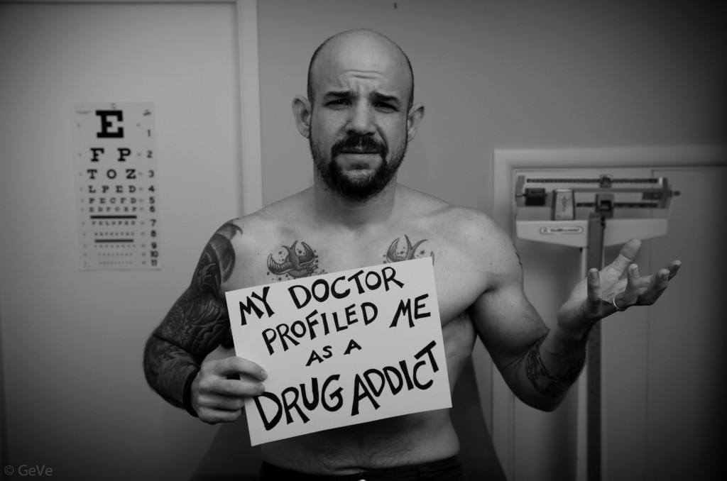
Patient Profiling: Drug Seekers, Medical Slang, and Malpractice

I posted an article that made me extremely angry. You can read it here:

ADDICTS USING GASTROPARESIS IN ORDER TO ABUSE PRESCRIPTION DRUGS.

I have heard story after story from people with chronic pain, invisible illnesses, and issues that illustrate why it is terrible for healthcare professionals to make snap judgments. They treat chronically ill patients as drug seekers. It can be life threatening if healthcare professionals just write someone off as a drug seeker without evaluating them. I just want to ask the doctors, "do drug addicts not have medical emergencies, too?" Patient profiling has become a REALLY big problem in our healthcare system, as has the "opioid epidemic."

I have written two articles on chronically ill patients being treated as drug addicts, and you can find them here:

GASTROPARESIS PATIENTS VS DRUG SEEKERS.

INVISIBLE EMERGENCIES SERIES PART THREE.


First of all, let me define patient profiling. According to Kevin MD,

"Patient profiling is the practice of regarding particular patients as more likely to have certain behaviors or illnesses based on their appearance, race, gender, financial status, or other observable characteristics. Profiling disproportionately impacts patients with chronic pain, mental illness, the uninsured, and patients of color. Like racial profiling by police, patient profiling by physicians is more common than you think.

We rely on doctors to first do no harm–to safeguard our health–but profiling patients often leads to improper medical care, and distrust of physicians and the health care system, with potential lifelong consequences."


Image Source: HERE


Can You Sue A Doctor For Patient Profiling?


According to Find Law,


"By Ephrat Livni, Esq. on April 01, 2016 3:55 PM

Patient profiling is a term used to describe a kind of discrimination by doctors. When a healthcare provider treats a patient based on their "type" rather than assessing them individually, that is profiling, and it can lead to problems in treatment.

Doctors should assess each patient individually, but profiling alone is not likely going to be a basis for a lawsuit against a doctor or hospital, unless that profiling manifested in medical malpractice. So let's explore the distinction between profiling, which is certainly unpleasant, and negligence law, which is based on actual injury.


Patient Profiling Primer

Dr. Pamela Wible, writing for Med Page Today's blog, Kevin MD, discusses and defines patient profiling. She explains, "Patient profiling is the practice of regarding particular patients as more likely to have certain behaviors or illnesses based on their appearance, race, gender, financial status, or other observable characteristics."

"Profiling disproportionately impacts patients with chronic pain, mental illness, the uninsured, and patients of color. Like racial profiling by police, patient profiling by physicians is more common than you think," Dr. Wible writes. "Profiling patients often leads to improper medical care."

That is where profiling starts to meet negligence in medicine. But there is still a distinction, as a medical malpractice suit must be predicated on an injury or harm that is compensable.


Medical Malpractice

Medical malpractice is a form of negligence for medical professionals. Negligence in medicine manifests in errors in diagnosis, treatment, or illness management, for example.

If any of these are the cause of an injury to the patient and it can be proven that the doctor's failures led to it, and that there were no unforeseeable intervening causes, then you have proven part of a claim. You must also show and that this harm can be compensated.

Now, if a doctor's negligence arose as a result of patient profiling, and you were harmed then the doctor's profiling, to the extent it can be shown, will help prove your malpractice claim. But profiling and negligence are not the same and one does not necessarily lead to the other, although it may be a strong sign that malpractice will arise if you are profiled as a patient.


Talk to a Lawyer

If you were treated poorly by a medical professional and were injured as a result, speak to a lawyer. Many personal injury attorneys consult for free or a minimal fee and will be happy to assess your claim."


Image Source: HERE


Failed/Erroneous Diagnosis and Treatment


"We all tend to trust our doctors. After all, they’re the experts. Even though we have some of the best doctors in the world, even they can make a mistake. And these mistakes can be a lack of treatment or the wrong treatment. These can be confusing concepts, so here is a brief overview on failed or erroneous diagnoses and treatments in medical malpractice cases.


Medical Misdiagnosis Generally

In most cases, if a delay or failure to diagnose a disease has resulted in injury or disease progression above and beyond that which would have resulted from a timely diagnosis, medical professionals could be held liable. Most doctors are trained to think and act by establishing a "differential diagnosis." Doing so calls for a doctor to list, in descending order of probability, his or her impressions or "differing" diagnoses of possible causes for a patient's presenting symptoms.

The key question in assessing a misdiagnosis for malpractice is to ask what diagnoses a reasonably prudent doctor, under similar circumstances, would have considered as potential causes for the patient's symptoms. If a doctor failed to consider the patient's true diagnosis on his/her differential diagnosis list, or listed it but failed to rule it out with additional tests or criteria, then the doctor is likely able to be sued for medical malpractice.

However, this situation may be difficult to prove. For example, a patient may allege that a doctor failed to timely diagnose a certain cancer, resulting in "metastasis" (spread of the cancer to other organs or tissues). But experts may testify that "micrometastasis" (spreading of the disease at the cellular level) may occur as much as ten years before a first tumor has been diagnosed, and cancerous cells may have already traveled in the bloodstream and lodged elsewhere, eventually to grow into new tumors. Therefore, it may be difficult in some cases to establish that a patient has suffered a worse prognosis because of the failure or delay in diagnosis.


Failure to Treat and Erroneous Treatment

If a patient is treated for a disease or condition that he or she does not have, the treatment or medication itself may cause harm to the patient. This is in addition to the harm caused by the actual condition continuing untreated.

The most common way in which doctors are negligent by failing to treat a medical condition is when they "dismiss" the presenting symptoms as temporary, minor, or otherwise not worthy of treatment. This situation may result in a worsening of the underlying condition or injury, causing further harm or injury. For example only, an undiagnosed splinter or chip in a broken bone may result in the lodging of a piece of bone in soft tissue or internal bleeding caused by the sharp edge of the splintered bone.

Erroneous treatment is most likely to occur as a result of a misdiagnosis. However, a doctor who has correctly diagnosed a disease or condition may nonetheless fail to properly treat it. Other times, negligence is the result of a doctor attempting a "novel" treatment that fails, when in fact a more conventional treatment would have been successful.


Getting Legal Help with a Medical Malpractice Claim

Having an illness or injury is bad enough, without thinking our doctors made it worse. And it’s not always easy to know whether the professionals in charge of your medical care acted properly. If you would like to know more about your claim, you can contact an experienced medical malpractice attorney.


Medical Malpractice In-Depth


When illness or injury forces you to see a physician or go to the hospital, you can generally be assured that a medical professional's years of experience and training will result in excellent treatment. But in truth, medical care providers are only human, and errors are always possible. Medical malpractice occurs when a negligent act or omission by a doctor or other medical professional results in damage or harm to a patient. To get started with a medical malpractice case, read First Steps in a Medical Malpractice Claim. See FindLaw's Medical Malpractice section for more articles and resources.


Negligence by a medical professional can include an error in diagnosis, treatment, or illness management. If such negligence results in injury to a patient, a legal case for medical malpractice can arise against:

The doctor, if his or her actions deviated from generally accepted standards of practice;
The hospital for improper care or inadequate training, such as problems with medications or sanitation;
Local, state or federal agencies that operate hospital facilities.


Medical malpractice laws are designed to protect patients' rights to pursue compensation if they are injured as a result of negligence. However, malpractice suits are often complex and costly to win. Therefore, if you believe you have a medical malpractice claim, it is important to consult with an attorney who will discuss your case with you, and help you determine your best options.


Legislation Affecting Malpractice Actions

Due in part to the power and resources of health care industry lobbyists, many states have passed legislation making it more difficult to bring and prevail in medical malpractice actions. In most states today, physicians and hospitals are protected by legal limits, called "caps," on the amount of damages and attorneys' fees that can be awarded in malpractice suits. Also, most states have a two-year time limit for filing malpractice actions, unless extraordinary circumstances affect the case.

One obstacle plaintiffs in many states may have to overcome before they can even file a malpractice action against a health care professional is the requirement that they file what is commonly known as a "certificate of merit." In order to file a certificate of merit, a plaintiff will first have to have an expert, usually another physician, review the relevant medical records and certify that the plaintiff's health care provider deviated from accepted medical practices, which resulted in injury to the plaintiff. The plaintiff's attorney then files the certificate of merit, which confirms that the attorney has consulted with a medical expert and that the plaintiff's action has merit.


"Respondeat Superior" and Independent Contractors

Medical malpractice can be committed by several types of health care professionals and, in a case where a hospital employee commits malpractice, the hospital itself may be held liable under the legal doctrine of "respondeat superior." Under this theory, an employer may be held liable for the negligent acts of its employee if the employee was acting within the scope of his or her employment when the negligence occurred. This doctrine is very important to plaintiffs in medical malpractice cases, because it helps ensure there will be a financially responsible party to compensate an injured plaintiff.

In some situations, commonly involving attending physicians working in hospitals, health care providers are considered independent contractors rather than employees, which makes the doctrine of "respondeat superior" inapplicable. What this means is, if a doctor or other health care professional an independent contractor, and commits malpractice while treating a patient in a hospital, the hospital cannot be held liable for the doctor's negligence. However, the hospital can be held liable for its own negligence, for example, in granting attending privileges to an unlicensed or incompetent physician.


Seek Legal Help with a Medical Malpractice Attorney

It's not always easy to know how to pursue a medical malpractice case. A qualified medical malpractice attorney will be able to discuss the strengths and weaknesses of your case and help you get the compensation you deserve. A good first step is to contact a medical malpractice attorney."


Image Source: On Image. Doctors are scared to prescribe pain medications due to the rising number of deaths and pressure from above


According to Parent Professional,


"Have you ever heard of patient profiling? It takes place when medical–and mental health–professionals make an assumption about someone seeking care based on their appearance, race, gender, financial status or even the kind of illness they have, such as mental health or substance use problems. The first time I came across this was in an article written by Pamela Wible, MD (article below at the end of this article), who recounted patient stories where the personal judgment of a medical person resulted in poorer care. She worried that, similar to racial profiling by police, patient profiling is more common than we want to admit. And it undermines care.

When I first heard about patient profiling, I immediately thought of my younger son. A few years ago (when he was in his early 20s), he woke up on a Sunday morning with horrible vertigo, He couldn’t stand, couldn’t focus and couldn’t drive. I took him to the local emergency room where they asked a series of routine questions: Are you on any medication? (No.) Have you ever experienced this before? (No.) Have you had anything alcoholic to drink? (Yes, one beer last night with friends. I was the designated driver.) Unfortunately, the questions stopped after he said he had had that beer the night before. He was given intravenous fluids, allowed to rest and sent home. The next day, his very irate primary care doctor sent him to a different emergency room where he was treated for inflammation of the inner ear.

For my son, staff at the first emergency room decided that a young adult in his 20s experienced vertigo because he had been drinking. They made a snap judgment and his treatment was delayed. To this day, he feels a general mistrust of emergency room staff.

For children and youth with mental health needs and their families, patient profiling happens far too often. It happens in the emergency room and it happens in visits to medical specialists. One mom, whose daughter had both a diagnosis of depression and frequent migraines – for which she was seeing a specialist – waited four days recently in the emergency room because no inpatient beds were available. She was told that her daughter couldn’t receive migraine medication while waiting because that was drug seeking behavior And it was probably part of the bipolar anyway. The mother was frantic when she called us and very frustrated that her daughter’s care was all being lumped under mental health. She felt the emergency room staff had stopped their assessment of her daughter’s needs after they heard about the bipolar disorder.

This doesn’t just happen in emergency rooms. It happens with medical specialists who think that mental health concerns have caused medical symptoms. It happens when doctors call parents 'enmeshed' or 'co-dependent' and don’t see them as a resource and partner but instead as part of the problem. It happens when young people are seen as their diagnosis and not as a valuable self-reporter and critical thinker.

That said, there is a fine line between patient profiling that can help or harm. Doctors, nurses, therapists and other workers often form an initial impression based on their experiences or their training. They often need this starting point to determine a course of action. But – and this is the crucial piece – that starting point needs updating as new information comes in. A second impression or a third is often in order. When the initial judgment is incomplete or inaccurate and it is not revised, it can be harmful.

A cornerstone of good care is excellent communication. While this is often characterized as the doctor or medical professional communicating to the patient, it should be a two-way street. Mutual exchange of information is critical but so is mutual listening. In any human interaction, the only way we can truly connect is when we get past our snap judgments and see who is actually there.

When a child is in crisis or when her need for care is urgent, parents are rarely at their best. Most often, there have been many stressful days or weeks before this point which have worn them down. We rely on medical staff to see beyond the diagnosis to the whole child, teen or young adult. We trust them to see our commitment and strength in the midst of the frenzy. We hope they will see us as a key member of the team, not as a 'less than' parent to be held at arm’s length.

When this doesn’t happen due to patient profiling, we all lose."


Image Source: HERE


According to the Daily Mail,


"Medical jargon is pretty impossible for most patients to follow, but some of the terms you hear your doctor use may just be insulting industry jargon.

Over decades, doctors have ad-libbed a whole vocabulary to encode their frustrations with problem patients, communicate grim status updates, or even gossip about children.

In medicine, this slang is more than harmless insider-jargon: studies have shown that doctors' attitudes and discrimination toward patients can have a real affect on the treatment they receive and how well they recover.

Medical schools have begun to recruit more diverse students in the hopes of changing the field's culture, but several young doctors who wished to remain anonymous told Daily Mail Online that discriminatory terms are still common.

But discrimination is institutionalized and dangerous in medicine, according to Dr Peter Muennig and Dr Alex Green of Harvard University and Massachusetts General Hospital, and it's worth knowing some of these terms that doctors may use to mask the severity of a situation or downright insult you (warning: some of these are offensive).


GOMER

This 'classic' term stands for 'get [them] out of my emergency room.' It has been used in hospitals for decades and is familiar to just about every doctor working, Dr Muennig says.

The acronym is a sort of catch-all term for any of the kinds of patients doctors don't want to deal with.

'There's a certain level of discrimination against the chronically ill, and that's where GOMER comes from,' says Dr Muennig.

In a recent interview with Daily Mail Online, he also said that this term is often used particularly to describe people who physicians suspect are hunting for pain medication.


Frequent fliers

Patients may return to the hospital week after week - or even day after day - for a variety of legitimate or illegitimate reasons, earning them the title 'frequent fliers,' but certainly no points from doctors.

Chronically ill patients with conditions like diabetes must make regular appointments for dialysis.

Other patients become common faces in emergency rooms and clinics because of their hypochondriacs tendencies, constantly sure that they are gravely ill.

Still other patients may just be looking for a drug fix, coming in with complaints of chronic pain, or in hopes that the physicians on shift will be more willing to prescribe than yesterday's were.

'It's most often used to talk about people with severe diseases like diabetes, or renal failure and diabetes,' says Dr Muennig.


FLK

Doctors use this cruel acronym for 'funny looking kid' to describe 'those babies who are "syndromic" or [we can tell] something is wrong with them based on how they look,' a Chicago doctor told Daily Mail Online.

Children born with any of the three trisomies - genetic mutations that cause there to be an extra copy or partial copy of a chromosome - often have distinct appearances.

These disorders include Down syndrome, which is typified by a flattened face, smaller head and ears than normal and upward slanting eyes.

Doctors also use the FLK to describe babies that don't have a clear diagnosis, but whose 'abnormal' appearance suggests that there may be something wrong with them.


Crumping

Not to be confused with the dance style, doctors use 'crumping' when they have a patient that is 'crashing, but not aggressively,' the Chicago doctor told Daily Mail Online.

The phrase is synonymous to 'circling the drain.' Rather than their organs suddenly failing - or crashing - these patients are deteriorating quickly, and often don't have much chance for survival.

'I don't think it's necessarily harmful to say "crumping" or "frequent flier,"' a New York-based doctor said, 'but I'm very careful as a physician to not use those words in earshot of a family, that would be strongly unprofessional.'


Total body dolores

Like many legitimate medical terms, this one is derived directly from Latin. 'Dolores' translates to pain so this 'literally means total body pain,' the doctor says.

The phrase is most often used between doctors, to describe a patient, as in, 'I have a total body dolores in room 109.'

He says he's seen this term used commonly to describe Latino patients in particularly. 'These patients can be very nervous,' he says, in part because English is often their second language, making the hospital and medical-speak even more frightening, 'and their anxiety manifests physically.'

Slang is used 'commonly with people of color, people using drugs,' he says. More common phrases like '"crack head" are commonly heard on the ward too.'

Although 'doloroes' means pain in Latin, a rather medical language, it means the same thing in Spanish, as well as being a woman' name. This all adds up to maximize confusion for patients.


Status dramaticus

A patient earns the title 'status dramaticus' when they are a '10/10 [for pain] always, although they look fine,' the Chicago doctor says.

'This is someone that wails shrieks, howls so loudly you can hear them from the hallways. Everything hurts and they make sure you know about it,' he says.

The problem with doctors using terms like status dramaticus, says Dr Alex Green, 'is that they're dealing with people who are sick, physically and sometimes mentally and these [dismissals] can be more directly harmful.'


WWWS

The acronym for 'wealthy white woman syndrom ' is 'actually bad because it's a term that is used when you're frustrated at specific patients and dismissing their symptoms,' says Dr Lisa Wang, a psychiatry resident in New York.

Research has documented that physicians are more likely to take women's pain less seriously than they would a man's. There is a widely held belief - though studies turn up mixed results - that women's bodies are designed for childbirth and their pain thresholds are higher.

A 2008 study from the National Institutes of Health also found that women wait 16 minutes longer to be seen in an emergency room than men do.

'As medical providers, on an unspoken level, I think [using these terms] is a coping mechanism, to make light of really difficult situations,' Dr Wang says.


HHS and Aye-aye-itis

Discrimination against people of color and those for whom English is a second language is rampant, as evidenced by the terms 'hysterical Hispanic syndrome' and aye-aye-itis.

'I hear "aye-aye-itis" used for a Hispanic patient who has many somatic complaints, none of which are related to their surgery or main problem, usually accompanied by "aye, aye,"' the Chicago doctor says.

'Language barriers are a big discriminatory factor,' says Dr Green.

'There's an attitude of "oh, another patient that doesn't speak English, why don't they learn, it makes our lives so difficult,' says Dr Green, who has worked on initiatives to educate hospitals on language barriers and introduce interpreters.

'I roll my eyes every time I hear that [kind of term]' says a New York doctor. 'I call it out because it gets at the rampant cynicism to the job.'

In one Harvard study, 20 percent of a group of 8,000 Latino people reported experiencing discrimination at a health care facility or clinic.


Slugging

'A slug is someone who is reluctant to get up out of bed after surgery,' says the Chicago doctor.

'They tend to be slow, in pain, and want to stay "one more day longer, please,"' he says.

On one hand, 'poor effort,' as he calls it, can have a negative impact on the quality and timeline for recovery, but there is a darker side to this tendency too.

A hospital bed might be the safest and most certain place some patients can stay, especially for those who are not financially secure or may be struggling with addiction.

'When you have patients showing up drunk every single night, always overdosing, not taking their blood pressure medications and coming in because of it, it becomes easy to blame patients, though I don't think you should,' says one New York doctor.

Dr Green says that 'a lot of it derives from [doctors'] systemic frustrations with the medical system back-firing back onto patients.'"


I have written a companion article with ALL of the medical slang, acronyms, etc. all on its own, because there is SO much of it. You can find the article: http://www.emilysstomach.com/2019/04/patient-profiling-medical-slang-only.html


Image Source: HERE


According to Pamela Wible MD,


"Patient Profiling: Are You a Victim?


We rely on doctors to first do no harm–to safeguard our health–but profiling patients often leads to improper medical care, and distrust of physicians and the health care system, with potential lifelong consequences. For the first time, people share their stories:

'I was once denied pain meds after a fall off a 10-foot porch by the same doc who gave my pretty female friend pain meds after getting two stitches in her finger. I felt like my appearance had something to do with it.' ~ Jay Snider

'In 1986 I was in a motorcycle accident. I tore up my face on the road. I was taken to the ER and treated like crap because I had no insurance. They cauterized my facial wounds rather than stitch me up, and then dumped me on the sidewalk with amnesia. I still have distinct black scars; people think they’re tattoos. I went into collections and it took years to pay that one off. Six weeks ago, I fell while trimming a tree. When the ER found the insurance card in my wallet, I was treated like gold.' ~ James Cummings

'As a teen, I fractured my nose. Many sinus issues later, I consulted an ENT specialist. He insisted that I damaged my sinus passages by using cocaine. His assumptions caused me pain, humiliation, confusion, and anger. I repeatedly assured him that I wasn’t a user. Two surgeries later, my septum was removed. Afterwards, he was so cruel as to continue his tirade about my cocaine use. As the gauze was being removed from my nose, I fainted. When I was roused, he insisted that I leave immediately showing no concern about whether I could even make it home safely.' ~ Lonnie Stoner

'It was 1975. I was 23 and I’d been on the pill for 4 years, but I became concerned about potential negative side effects of long-term hormonal manipulation. So I researched other contraceptives and felt the diaphragm was the simplest and safest option for me. When I went to the county clinic to get fitted, I explained what I’d researched to the doctor. He scoffed at my concerns, urged me to stay on the pill, and disputed any potential negative consequences. He reminded me that taking a pill each day was SO much easier than having to be responsible for using the diaphragm properly. It was clear he thought I was too young and clueless to make this decision about my own reproductive health care. Although he tried to dissuade me from switching to a diaphragm, I insisted that’s what I wanted, and he finally fitted me for it. After he left the room, the nurse said, ‘Don’t worry, dear; it’s quite easy to use. I’ve been using one for years with no problems. It’s a good choice for you to make!’ It was clear she didn’t approve of his patronizing attitude either.' ~ Patsy Raney

'I injured my back at work. I couldn’t get time off, so my family doc prescribed pain meds so I could get through the day and Xanax for sleep. I returned every six months for two years and he always accused me of taking more than I was prescribed. He got progressively more rude and angry. I brought my wife with me to see if I was imagining it. She witnessed it too, so we searched for another doctor. I asked my new doctor to taper me off of the pain meds and Xanax so I could try medical marijuana instead. He was skeptical. He told me to go to the pain clinic. I’d gone there once before and was treated like a criminal. I didn’t want to go there! So he wrote up a contract that said I would agree to take pain meds and Xanax and I’d be drug tested monthly to make sure that I wasn’t using medical marijuana. When I told him I wouldn’t sign the contract, he told me to find another doctor. This was at a critical time when I needed real help and was worried about taking the meds for over two years.' ~ Carl Williams

I’ve been a doctor for 20 years. I thought I’d seen it all. Drug addicts have altered my prescriptions, even forged my name. Patients have lied to me. Many haven’t followed my treatment plans. Some have died as a result. Still, I try to treat everyone fairly and with respect. But now I’m wondering, “Have I ever profiled a patient?” I bet I have. So on behalf of my colleagues and myself, I’ve got a message for any patient who has ever been misjudged or mistreated:


Image Source: In Article


Special shout out to Chriss for her help.


Monday, March 11, 2019

Weight Gain, Starvation, & Gastroparesis

A friend of mine sent me a message about two months ago asking me questions about Gastroparesis because she was scared. The Gastroenterologist she saw told her a lot of different things that did not sound right to her, so she questioned it. I am very glad she did because he gave her a lot of misinformation that if she would have listened to, she might have died. The doctor she saw for her Gastroparesis told her she was too overweight for him to treat her.

He suggested that she go on a diet and starve herself a bit before he would consider treating her. She told him that she was starving and that her body was starving itself because she was unable to keep down food, which is why she went to the doctor. He told her that "starvation mode" was not a real thing. I was flabbergasted when she was relaying to me what he said. Additionally, she is also diabetic, so she cannot exactly "starve" herself (There is a lot of controversy regarding whether or not "starvation mode" is real. There are arguments on both sides but I will write about it, with arguments from both sides, and share sources, for you to make up your own minds).

I told her to see another doctor immediately because I know "starvation mode" is a real thing. My own doctor has told me that my body is starving itself from all of the vomiting and barely eating. I came home and did research immediately.

Some medical professionals see that patients are overweight and because they have this preconceived notion of what Gastroparesis is supposed to look like, they do not take the heavier Gastroparesis warriors seriously. I want to clarify that people can gain weight with Gastroparesis and they can also lose weight with it, too.

These are both complex issues with complex answers. I am glad my friend reached out to me to ask questions instead of following her doctor's advice to the letter, because I really do not know what would have happened to her if she had. I am so glad she was curious and smart enough to question the advice she was told, and went to get a second opinion. Sadly, this is not the first time I have heard this story. I have had a lot of people tell me that their doctors wanted them to lose weight first, before they would treat them, and/or some alternate versions of they did not need a feeding tube because they were too overweight for one. Let me make this VERY clear, your weight has nothing to do with malnutrition, vitamin deficiencies, or anything like that. I really hate it when medical professionals bring up weight, because it really sounds demeaning, patronizing, and I feel like I am being judged.


I made a survey to ask people with Gastroparesis whether or not they were treated differently because of their weight. I received sixty-three responses, which is amazing. They can be found HERE. To summarize the surveys I received, basically some doctors (not all), friends, and family members do not believe people have Gastroparesis if they are overweight. When you have Gastroparesis, you can gain weight or lose weight. Let's talk about weight gain first.**


“Eating too few calories, for too long of a period will cause your metabolism to slow down so much that it will prevent weight loss and even cause weight gain.”


Image Source: https://i.pinimg.com/originals/6f/21/6d/6f216d9733be5419e33ba262edde42fd.png


Weight gain can not only be attributed to starvation mode, where your metabolism slows, but could be caused by stress and anxiety in addition to your metabolism rate: http://www.emilysstomach.com/2017/01/mental-health-and-gastroparesis-part-1.html


Some of these sources refer to weight gain and starvation mode for dieting for healthy people, but it can be applied to Gastroparesis as well, since we do not have the luxury of eating to consider a diet.


Source: https://tinyurl.com/yapse9lm


Starvation is a severe deficiency in caloric energy intake. It is the most extreme form of malnutrition. In humans, prolonged starvation can cause permanent organ damage and eventually, death, according to the World Health Organization. The bloated stomach represents a form of malnutrition called kwashiorkor which is caused by insufficient protein despite a sufficient caloric intake.


My article called, "What Gastroparesis Does to the Body" that you can find HERE.

My article on Vitamin Deficiencies can be found HERE.


According to MedLine Plus,


"Kwashiorkor is a form of malnutrition that occurs when there is not enough protein in the diet.


Causes:

Kwashiorkor is most common in areas where there is:

Famine
Limited food supply
Low levels of education (when people do not understand how to eat a proper diet)


This disease is more common in very poor countries. It often occurs during a drought or other natural disaster, or during political unrest. These conditions are responsible for a lack of food, which leads to malnutrition.

Kwashiorkor is rare in children in the United States. There are only isolated cases. However, one government estimate suggests that as many as 50% of elderly people in nursing homes in the United States do not get enough protein in their diet.

When kwashiorkor does occur in the United States, it is most often a sign of child abuse and severe neglect.


Symptoms

Symptoms include:

Changes in skin pigment
Decreased muscle mass
Diarrhea
Failure to gain weight and grow
Fatigue
Hair changes (change in color or texture)
Increased and more severe infections due to damaged immune system
Irritability
Large belly that sticks out (protrudes)
Lethargy or apathy
Loss of muscle mass
Rash (dermatitis)
Shock (late stage)
Swelling (edema)


Exams and Tests

The physical exam may show an enlarged liver (hepatomegaly) and general swelling.

Tests may include:

Arterial blood gas
BUN
Complete blood count (CBC)
Creatinine clearance
Serum creatinine
Serum potassium
Total protein levels
Urinalysis


Treatment

Getting more calories and protein will correct kwashiorkor, if treatment is started early enough. However, children who have had this condition will never reach their full potential for height and growth.

Treatment depends on the severity of the condition. People who are in shock need treatment right away to restore blood volume and maintain blood pressure.

Calories are given first in the form of carbohydrates, simple sugars, and fats. Proteins are started after other sources of calories have already provided energy. Vitamin and mineral supplements are essential.

Since the person will have been without much food for a long period of time, eating can cause problems, especially if the calories are too high at first. Food must be reintroduced slowly.

Many malnourished children will develop intolerance to milk sugar (lactose intolerance). They will need to be given supplements with the enzyme lactase so that they can tolerate milk products.


Outlook (Prognosis)

Getting treatment early generally leads to good results. Treating kwashiorkor in its late stages will improve the child's general health. However, the child may be left with permanent physical and mental problems. If treatment is not given or comes too late, this condition is life threatening.


Possible Complications

Complications may include:

Coma
Permanent mental and physical disability
Shock


When to Contact a Medical Professional

Call your health care provider if your child has symptoms of kwashiorkor.


Prevention

To prevent kwashiorkor, make sure the diet has enough carbohydrates, fat (at least 10% of total calories), and protein (12% of total calories).


Alternative Names

Protein malnutrition; Protein-calorie malnutrition; Malignant malnutrition


Kwashiorkor is a condition resulting from inadequate protein intake. Early symptoms include fatigue, irritability, and lethargy. As protein deprivation continues, one sees growth failure, loss of muscle mass, generalized swelling (edema), and decreased immunity. A large, protuberant belly is common. The incidence of kwashiorkor in children in the United States is extremely small and it is typically found in countries where there is drought and famine.


Image Source: In the Article


References

Ashworth A. Nutrition, food security, and health. In: Kliegman RM, Stanton BF, St. Geme JW, Schor NF, eds. Nelson Textbook of Pediatrics. 20th ed. Philadelphia, PA: Elsevier; 2016:chap 46.

Kumar V, Abbas AK, Aster JC. Environmental and nutritional diseases. In: Kumar V, Abbas AK, Aster JC, eds. Robbins and Cotran Pathologic Basis of Disease. 9th ed. Philadelphia, PA: Elsevier Saunders; 2015:chap 9.

Manary MJ, Trehan I. Protein-energy malnutrition. In: Goldman L, Schafer AI, eds. Goldman-Cecil Medicine. 25th ed. Philadelphia, PA: Elsevier Saunders; 2016:chap 215.
Review Date 2/19/2018

Updated by: Neil K. Kaneshiro, MD, MHA, Clinical Professor of Pediatrics, University of Washington School of Medicine, Seattle, WA. Also reviewed by David Zieve, MD, MHA, Medical Director, Brenda Conaway, Editorial Director, and the A.D.A.M. Editorial team."


Image Source: https://62e528761d0685343e1c-f3d1b99a743ffa4142d9d7f1978d9686.ssl.cf2.rackcdn.com/files/88935/area14mp/image-20150720-21056-kw51zj.png


Image Source: https://www.google.com/url?sa=i&rct=j&q=&esrc=s&source=images&cd=&cad=rja&uact=8&ved=2ahUKEwjd-__GlfvgAhUlgK0KHcmTAIIQjRx6BAgBEAU&url=http%3A%2F%2Ftheconversation.com%2Fchemical-messengers-how-hormones-make-us-feel-hungry-and-full-35545&psig=AOvVaw3woiWmpXezgj7-o0y1ytU7&ust=1552430364356055


Image Source: https://www.healthline.com/health/low-blood-sugar-effects-on-body#1


I had no idea there was a hunger scale, like the pain scale, until I started doing research for this article.


According to Medical News Today,

"Malnutrition results from a poor diet or a lack of food. It happens when the intake of nutrients or energy is too high, too low, or poorly balanced. Undernutrition can lead to delayed growth or wasting, while a diet that provides too much food, but not necessarily balanced, leads to obesity. In many parts of the world, undernutrition results from a lack of food. In some cases, however, undernourishment may stem from a health condition, such as an eating disorder or a chronic illness that prevents the person from absorbing nutrients."


According to Fitness,

"Starvation is dangerous to deal with and leads to all sorts of problems, both mental and physical. Want to know what they are? Read this post and find out about the harmful and potentially dangerous side effects of starving yourself.

Starvation is a condition that is often accompanied by severe deficiency in your calorie intake. Starvation can be self-inflicted (hardcore dieters) or due to the presence of other conditions like anorexia or bulimia. Starvation can also lead to severe malnutrition and may permanently damage your internal organs.

Starvation isn’t something pleasant, and you should try and refrain from starving yourself, especially under the pretext of following a diet regime. To discourage you further, let’s look at some of the adverse effects of starvation in detail:


1. MALNUTRITION:

This is perhaps the most damaging effect of voluntary or forced starvation. Malnutrition is a terrible condition, and it affects most children in developing countries. Starvation usually means that you don’t eat much at all. This leads to many essential nutrients and vitamins being excluded from your diet. Malnutrition can lead to various mineral and nutrient deficiencies like Night blindness (vitamin A deficiency), Scurvy (Vitamin C deficiency) and even Anemia (Iron deficiency) (2).


2. DEHYDRATION:

The second most obvious result of starvation; just like a lack of food induces malnutrition, a lack of water can induce dehydration. This dehydration may in turn aggravate other problems and lead to cracked and exceedingly dry skin, along with other conditions like constipation, among others. Thus, it is important to remember to drink enough water, even if you are on a starvation diet. Starvation and dehydration depletes glycogen levels and disturbs electrolyte balance (3).


3. Decreased Resting Metabolic Rate:

Starvation can lead to a reduced resting metabolic rate. Your Basal metabolic rate or BMR is defined as the minimal rate of energy expenditure per time unit while you rest. Tests found that fasting prior to rest can lead to an increase in white adipose tissue (WAT) and decrease in flow to the brain. Other studies determine that starvation can induce a decrease in your basal metabolic rate.


4. Loss Of Monthly Menstrual Periods:

Starvation leads to many problems. One of these problems is it upsets how your periods are regulated. So, for those of you thinking about starvation diets, you might want to consider this before you begin. In some cases, if you are too underweight your body may simply cease to produce estrogen. A lack of fat can restrict your cells from converting cholesterol to extra estrogen. As starvation can lead to organ damage, body processes tend to slow down, which usually causes menstruation to stop (4).


5. Constipation:

Starvation can also induce constipation in many individuals. Studies on many anorexic individuals conclude that people who suffer from the disorder may suffer from severe starvation, which can lead to many disorders and requires the use of regular laxatives to be treated wholly. A case study on three severely anorexic women found that these women needed regular laxatives or enemas to help flush their colon. These women also ran a higher risk of suffering from rectal prolapse due to the overuse of laxatives (5).


6. Bone Loss:

A starved or starving individual may continue to lose weight at an alarming rate. Although the changes in your skin and musculature are quite obvious, but what about the changes in your bone structure! Did you know that starvation may lead to bone density loss? Well, usually when you consume a calorie-restricted diet, your bones are remodeled. The older bone breaks down and new bones form rapidly. However, your bone density reduces, which can lead to fragility (6).


7. Fatigue:

One of the most common side effects of starvation is fatigue. You have all heard about ‘eating to keep your strengths up,’ this idiom cannot ring any truer as eating a balanced diet helps provide energy for the body primarily, while helping keep diseases and infections at bay. Starvation upsets the balance of essential vitamins in your body and slows down physiological processes, which can also lead to fatigue and a feeling of faintness. Starvation is often associated with a feeling of lightheadedness (7).


8. High Blood Pressure:

Malnutrition and starvation can subject your body to many conditions, including high blood pressure. When you starve, essential nutrients like potassium and vitamin D aren’t consumed, which leads to a spike in blood pressure and many other deficiencies. All these factors ensure that starvation inevitably raises your blood pressure. In a study conducted on three groups of children, it was found that two groups: one malnourished and one recovering from malnourishment had significantly higher blood pressure readings than the children who weren’t malnourished at all.


9. Electrolyte Imbalance:

Starvation leads to a loss of nutrients, which results in the consumption of fewer electrolytes with each passing day. Heart-healthy Minerals like potassium, sodium, calcium and magnesium that promote proper heart function and heart health aren’t consumed when you don’t have enough food. Starving not only results in lesser nutrients, but also promotes electrolyte imbalance. Thus, remember to have mineral supplements if you are on a starvation diet.


10. Affects The Brain:

There have been many studies on how starvation can affect cognitive abilities and make you feel depressed. Try and recall how your friend snaps at you when she’s on a starvation diet. When we miss a meal, we often find ourselves dwelling on food and how it would feel to be properly fed.

You know that starvation can lead to many other physical symptoms like dehydration and fatigue that can impair your thinking abilities and also make you aggressive and jumpy. Starvation can also affect how you react to stress and can induce feelings of anxiety, irritability and even lead to chronic or clinical depression in some cases.

Now that you know how starvation can adversely affect your body, we hope that you will rethink about those starvation diets. They are just a quick fix and offer little long term benefit. Opt for a balanced regimen to lose weight."


Image Source: https://i.pinimg.com/564x/3a/31/c0/3a31c05ee329a6ee1ee8e7d0f7633b11.jpg


According to Health Line,


"Is 'Starvation Mode' Real or Imaginary? A Critical Look


Weight loss is generally seen as a positive thing. It can bring improved health, better looks and all sorts of benefits, both physical and mental. However, your brain doesn't necessarily see it that way. Your brain is more worried about keeping you from starving, making sure that you (and your genes) survive. When you lose a lot of weight, the body starts trying to conserve energy by reducing the amount of calories you burn. It also makes you feel hungrier, lazier and increases your cravings for food. This can cause you to stop losing weight, and may make you feel so miserable that you abandon your weight loss efforts and gain the weight back.

This phenomenon is often called 'starvation mode,' but is really just the brain's natural mechanism to protect you from starvation.


What Does 'Starvation Mode' Imply?

What people generally refer to as 'starvation mode' (and sometimes 'metabolic damage') is the body's natural response to long-term calorie restriction. It involves the body responding to reduced calorie intake by reducing calorie expenditure in an attempt to maintain energy balance and prevent starvation. This is a natural physiological response, and isn't really controversial. It is well accepted by scientists, and the technical term for it is 'adaptive thermogenesis.' I will use the term starvation mode in this article, although it really is a misnomer because true starvation is something that is almost completely irrelevant to most weight loss discussions. Starvation mode was a useful physiological response back in the day, but does more harm than good in the modern food environment where obesity runs rampant.


Calories In, Calories Out

Obesity is a disorder of excess energy accumulation. The body puts energy (calories) into its fat tissues, storing it for later use. If more calories enter the fat tissue than leave it, we gain fat. If more calories leave the fat tissue than enter it, we lose fat. This is fact. Pretty much all weight loss diets cause a reduction in calorie intake. Some by controlling calories directly (counting calories, weighing portions, etc), others by reducing appetite so that people eat fewer calories automatically. When this happens, calories leaving the fat tissue (calories out) become greater than the calories entering it (calories in). So we lose fat. However, the body doesn't see this in the same way as you do. In many cases, it sees this as the beginning of starvation. So the body fights back, doing everything it can to make you stop losing.

The body and brain can respond by making you hungrier (so you eat more, increasing calories in), but what is most relevant to this discussion here is what happens to the amount of calories you burn (calories out). Starvation mode implies that your body reduces calories out in an attempt to restore energy balance and stop you from losing any more weight, even in the face of continued calorie restriction. This phenomenon is very real, but whether this response is so powerful that it can prevent you from losing weight, or even start gaining despite continued calorie restriction, is not as clear.


What people refer to as 'starvation mode' is the body's natural response to long-term calorie restriction. It involves a reduction in the amount of calories your body burns, which can slow down weight loss. The amount of calories you burn can change.


The amount of calories you burn in a day can be roughly split into 4 parts:

Basal Metabolic Rate (BMR): The amount of calories your body uses to maintain vital functions, such as breathing, heart rate and brain function.

Thermic Effect of Food (TEF): The calories burned while digesting a meal. Usually about 10% of calorie intake.

Thermic Effect of Exercise (TEE): Calories burned during physical activity, such as exercise.

Non-Exercise Activity Thermogenesis (NEAT): Calories burned fidgeting, changing posture, etc. This is usually subconscious.

It involves a reduction in movement (both conscious and subconscious), and a major change in the function of the nervous system and various hormones.


There are several ways that the body burns calories. All of them can go down when you restrict calories for a long time. Studies Show That Calorie Restriction Can Reduce 'Calories Out.' Studies clearly show that weight loss reduces the amount of calories you burn. According to a large review study, this amounts to 5.8 calories per day, for each pound lost, or 12.8 calories per kilogram. What this means, is that if you were to lose 50 pounds, or 22.7 kilograms, your body would end up burning 290.5 fewer calories per day. The reduction in calorie expenditure can be much greater than what is predicted by changes in weight. For example, some studies show that losing and maintaining 10% of body weight can reduce calories burned by 15-25%. This is one of the reasons weight loss tends to slow down over time, and why it is so difficult to maintain a reduced weight. You may need to eat fewer calories for life! Keep in mind that it is possible that this metabolic 'slowdown' is even greater in some groups that have a hard time losing weight, such as postmenopausal women.


Muscle Mass Tends to Go Down

Another side effect of losing weight, is that muscle mass tends to go down. As you may know, muscle is metabolically active, and burns calories around the clock. However, the reduction in calorie expenditure is actually greater than can be explained by a reduction in muscle mass alone. The body becomes more efficient at doing work, so less energy than before is required to do the same amount of work. So calorie restriction makes you expend fewer calories for the physical activity (whether deliberate or subconscious) that you perform.

Weight loss and reduced calorie intake can lead to reduced burning of calories. On average, this amounts to about 5.8 calories per pound of lost body weight.


How to Avoid the Metabolic Slowdown

Keep in mind that your metabolism slowing down is simply a natural response to reduced calorie intake. Although some reduction in calorie burning may be inevitable, there are a number of things you can do to mitigate the effect. The single most effective thing you can do is resistance exercise.


Starvation mode is real, but it's not as powerful as some people think. It can make weight loss slow down over time, but it won't cause someone to gain weight despite restricting calories. It's also not an 'on and off' phenomenon, like some people seem to think. It's an entire spectrum of the body adapting to either increased or decreased calorie intake. Starvation mode is actually a terribly inaccurate term. Something like 'metabolic adaptation' or 'metabolic slowdown' would be much more appropriate. This is simply the body's natural physiological response to reduced calorie intake. Without it, humans would have become extinct thousands of years ago. Unfortunately, this protective response can cause more harm than good where overfeeding is a much, much greater threat to human health than starvation."


Let's talk more about starvation mode. I want to quote a couple of different sources, and they may say similar things, but I want to look at this closely since my friend's doctor said it wasn't a real thing or problem. I just do not believe that. My doctor, when I was in high school (when I first started vomiting and getting sick, but didn't know what was wrong) told me that my body was starving itself. He had to explain to me what that meant and at that time, I was sixteen and ninety pounds (I am 5'4"). He told me I had to supplement what I ate with Ensure, so I had to eat an entire meal and drink Ensure on top of it. I actually really started gaining weight in 2009, when I fell down a mountain and broke both of my knees. But, I digress, because that isn't the point.

The point is, I had a doctor confirm to me, personally, that starvation mode is VERY real and people's bodies handle it in different ways. My body was feeding off of itself, consuming my muscle mass and things like that. Starvation is VERY real and VERY dangerous. It is SO dangerous for medical professionals to tell anyone that.

You have to be your own advocate and research things that you may not understand, that you may have questions about, and/or even doubts about.

I do not want to disparage doctors in any way, but they do not know everything. They do their utter best to rule things out and to help you as much as they are able to, and there are REALLY great doctors out there who care, but there are also doctors who do not even know about Gastroparesis or the effects it can have on someone (and given as much knowledge as they have to retain, I do not fault them for this). I do not want to alienate any medical professionals out there, as I respect the job that you do. This article is mainly to help misunderstandings and to educate the medical professionals, hopefully, that told my diabetic friend with Gastroparesis, to lose some weight before he would treat her for Gastroparesis, and that 'starvation mode' did not exist. Well, I'm going to research it and prove it does.


Image Source: https://anabolicminds.com/forum/attachment.php?attachmentid=72947&d=1358175074


According to InBodyUSA,

"Research indicates people who used starvation diets for weight loss, eating 50% of their energy needs for three weeks, did decrease their body weight overall. However, they also reduced their lean muscle mass by 5%. If the state of starvation is maintained chronically, lean muscle mass and organ size are decreased by 20%.

Likewise, a study on mice found lean mass and lean muscle mass were sacrificed during starvation; however, body fat stores were relatively the same in mice on a control diet and obese mice on starvation diets.

Weight loss via starvation causes individuals to lose significant amounts of lean muscle mass and Lean Body Mass, which encompasses water, bones, organs, etc. Reducing the mass of your bones is problematic, as that decreases bone density and can make you more prone to injury. Conversely, increasing Lean Body Mass increases bone strength and density, a common concern for many Americans as they age.

One study using human participants indicated dropping significant amounts of calories from the diet lead to significant weight loss and decreased lean muscle mass. However, participants also gained back nearly all of the fat they lost, within 8 years.

This prompts the discussion and understanding of an important topic- metabolism. More so, chronic starvation leads to changes in metabolism. Metabolism and resting metabolic rate are directly linked to Lean Body Mass. A person with greater body mass will require more energy to function day to day, thus will have a larger basal metabolic rate (BMR). As weight decreases, so does BMR.

This means that there’s a certain number of calories necessary to maintain your lean mass. If you go below this number, your body will be forced to break down these muscle stores in order to create energy.

Starvation diets have far-reaching negative effects on the body. Starving to lose weight changes the metabolism, reduces lean muscle, reduces bone density, and decreases strength."


Image Source: https://www.sciencedaily.com/images/2017/01/170126081628_1_540x360.jpg


There is an interesting book about Starvation Mode by Leigh Peele (that is in ebook form, too). This is her research and take on Starvation Mode and the Effects and Symptoms,

"Different Effects and Symptoms of Starvation Mode

Starvation happens when the body is deprived of the essential nutrients it requires for proper function and survival. When the body does not receive these nutrients that come from food and liquids, side effects occur. One of the effects of starve mode is weight loss and many people use this method to lose unwanted weight. This is often done as an act of desperation because of stubborn weight that they are unable to lose with all other weight loss techniques they have tried in the past. Going on starvation mode is not recommended to be done often since it can have irreversible effects that could eventually lead to death.

On starvation mode, the body feeds itself with what is inside it. Because you take in only a few calories, you lose weight. It should, however, be noted that when few calories enter the body, the metabolism also slows down. Because the metabolism burns calories, it burns only a few calories once it slows down. The moment you start consuming food regularly, the weight will come back. This is because the body begins to comprehend that it is starving and so it shuts down your weight loss efforts for it to survive.

Some of the bad effects of starvation mode include failed diets and other mental and physical ailments. These results are brought about by the malnourishment and depression. Eventually you will gain the weight that you lost back due to your coping mechanisms when you are depressed.

Starvation obviously makes you lose weight. The body does not have any sustenance inside it. You can lose up to two pounds a day with starvation. While it may initially sound good, the effects of such drastic weight loss might result in a medical emergency. Your body and your organs might not be able to cope with not having sustenance for days at length.

Starvation can cause dehydration because the body lacks fluid it needs. It then uses water and fluids already stored in your body. The first organ to be affected is the kidneys and they shall fail and eventually stop working once your urine output stops. Your skin will lose rigidity and turgor and shall become extremely dry and stiff. When you become dehydrated, your heart rhythms become irregular and this shall lead to heart diseases.

Another one of the effects of starvation mode is electrolyte imbalance. Because there is loss of fluids and nutrients, there is no fuel available for the body to work properly. Electrolytes make the heart, nerve impulse and muscle impulse function properly. They also make oxygen flow steadily in the body. If the flow is disrupted severely, you may slip into a coma. Blood sugar levels quickly drop because of the absence of glucose even if the pancreas produces just a small amount of insulin.

During the ultimate stage of starvation, severe muscle atrophy happens. The muscle crumbles and lessens because the body feeds from itself. It uses its muscle mass so as to give energy to the bloodstream so that the organs will not shut down. Muscle spasms and twitches happen when the potassium level becomes dangerously low. Extreme weakness and paralysis may also happen. When the muscles break down, the nerve cells deteriorate, especially those in your spinal cord area which regulates movement. If the muscles are weak and do not function, you may not move freely at all.

Low blood pressure or hypotension is also a side effect of starvation mode. Aside from such bouts, the temperature of the body also drops. When the blood pressure becomes so low, shock may happen and this is a very dangerous condition because the body shots down just to survive. If hypotension is left untreated, death or coma will happen."


Gastroparesis can effect everyone differently. Everyone's bodies are different and so Gastroparesis is harder to treat because it varies so widely from person to person. I wrote about this in detail, and if you would like to read that article, you can find it here: http://www.emilysstomach.com/2018/04/gastroparesis-different-ways-it-effects.html

For the many different causes of Gastroparesis and the available treatment options, you can read here: http://www.emilysstomach.com/2018/10/the-many-causes-of-gastroparesis.html

I also do not want anyone to think that Gastroparesis is an eating disorder. It is not. It can be caused by an eating disorder, but Gastroparesis, itself, is a chronic, invisible illness. For specifics on the differences between the two, please read this article: http://www.emilysstomach.com/2015/04/gastroparesis-vs-eating-disorders.html


More sources can be found at:

NDTV.

SIGN OF THE TIMES.

LIVESTRONG.COM. "A lack of food can cause the body to go into starvation mode over time. Starvation mode is a metabolic response to the body being deprived of food, which may occur during periods of famine or economic depression, when using a fad diet, or when suffering from anorexia nervosa. A variety of specific signs and symptoms affect those whose body has gone into starvation mode."


SOURCE:KHAN ACADEMY.


SOURCE: https://www.youtube.com/watch?v=eZNu5bsCyEc


THE CONVERSATION.


**You can click on the all capitalized bold words for additional information
Creative Commons License
This work is licensed under a Creative Commons Attribution-NonCommercial-NoDerivatives 4.0 International License.

Sunday, December 30, 2018

No Evidence of Impaired Gastric Emptying in Early Huntington‘s Disease

According to the PLOS Currents Huntington,


"No Evidence of Impaired Gastric Emptying in Early Huntington‘s Disease
November 16, 2011 · Epidemiology


Authors

Carsten Saft
Jürgen Andrich
Marc Fälker
Sarah Gauda
Sina Küchler
Dirk Woitalla
Oliver Goetze


Abstract

Background: Several factors, such as dysphagia, an increased motor activity, increased metabolic rate and a hypermetabolic state have been discussed as contributing to weight loss even at the early stages of Huntington’s Disease (HD). Aim of this pilot study was to investigate gastric emptying as a possible reason for weight loss in HD.

Methods: 11 HD participants at early stages of the disease and matched controls were investigated by using the well-established and non-invasive 13C-octanoate breath test. The “Gastroparesis Cardinal Symptom Index” and the “Short-Form Leeds Dyspepsia Questionnaire” were used for clinical evaluation of gastroparesis or dyspepsia.

Results: When compared to standard values ​​given in literature and controls all HD patients had normal breath test results. There was no evidence of gastroparesis or dyspepsia. There was a correlation of breath test results with the cognitive and functional performance of HD participants.

Conclusion: According to our data, there is no evidence of impaired gastric emptying in early HD. We can not exclude that gastric emptying contributes to weight loss at more advanced stages of the disease.

Corresponding author: PD Dr. med. Carsten Saft, Department of Neurology, Huntington-Center NRW, St. Josef Hospital, Gudrunstrasse 56, 44791 Bochum, Germany, E-mail: carsten.saft@ruhr-uni-bochum.de

§ Carsten Saft and Jürgen Andrich contributed equally to this work
Funding Statement
The study was supported by a FoRUM grant, University of Bochum (AZ: F506-2006). Oliver Götze was supported by the DFG (Gö 13582/1).
Introduction

Weight loss is a main feature in Huntington’s disease (HD) and was found to be manifest even at early stages of the disease. [1][2][3][4][5] Multifactorial causes, such as decreased caloric intake due to dysphagia and a higher energy expenditure due to increased motor activity have been discussed as being a possible reason for weight loss especially at the advanced stages of the disease. [6][7][8][9][10] Using a whole body indirect calorimetry in both early stage HD patients and the R6/2 transgenic mouse model of HD, Goodman and colleagues were able to demonstrate that patients with early HD tended to have a negative energy balance for reasons not related to their movement disorder, which was paralleled in the transgenic R6/2 mice. [4] This leads to the assumption of an increased metabolic rate as a main reason for weight loss in HD, which is supported by other experiments in the transgenic R6/2 mice. [4][11][12] In a study investigating the direct relation between the number of CAG repeats in the mutant huntingtin gene and weight loss, Aziz and colleagues found a correlation between both of these factors and discussed a hypermetabolic state as being a reason for weight loss, occurring even at early stages of the disease. [13] They discussed a hypermetabolic state as being likely to stem directly from interference of the mutant protein with cellular energy homeostasis and thus reflecting fundamental pathologic mechanisms underlying HD and not to be secondary to hyperactivity. Since mutant Huntingtin (mtHtt) is not only expressed in the brain of HD patients, but also in the gastrointestinal (GI) tract, a recently published study investigated the GI tract in the R6/2 mice model for HD. This study describes a loss of enteric neuropepitdes, a decreased mucosal thickness and villius length and also an impaired gut motility, diarrhea, and malabsorption of food, suggesting that GI dysfunction plays an important role in weight loss in HD mice. [14]

In addition, gastrointestinal dysfunction is discussed as being the main reason for weight loss in Parkinson’s disease (PD). [15] In a study using a solid meal and the 13 C-sodium octanoate breath test for measurement of gastric emptying in patients with PD, Goetze and colleagues found 88% of PD patients suufered from delayed gastric emptying when compared with controls. The severity of motor impairment was associated with gastroparesis. [16] Several other studies confirm an impaired gastric emptying in PD, some of them with a rate of 100% of PD patients. [16][17][18][19][20][21] One study describes a 60% delay in gastric half emptying time in the PD patient group after a solid test meal using the non-invasive 13 C-sodium octanoate breath test for evaluation of gastric emptying. [17] Neuropathological findings suggest enteric dysfunction to be one of the initial pathophysiological events in PD. [16][22] Central and enteric nervous system involvement in PD is discussed as being a pathophysiologic basis for this dysfunction. [15]

Autonomic nervous dysfunction was found to be present in HD, too. [23] Thus, the aim of the current study was to investigate gastric emptying in early HD patients without medication as a possible additional reason for weight loss by using the well-established 13 C-octanoate breath test. [16][17][18][19][20][21][24]
Methods


Participants

11 manifest HD patients with genetically confirmed diagnosis and without any medication in at clinically early stages of the disease (Shoulson stage I/II) and 11 controls were recruited from the HD centre Bochum, Germany. [25] Participants with known concurrent gastrointestinal diseases or previous operations of the gastro-intestinal tract were excluded, as well as patients with other severe diseases, diabetes mellitus, severe respiratory dysfunction, and malignancies. Also participants with concurrent liver diseases or excessive alcohol consumption (50 g/d of ethanol) were excluded. All participants had lab parameters for ALT, AST, LDH, cholesterol and triglycerides within the normal range, as well as normal findings for the ultrasonography of the upper abdomen. Pregnant and breast-feeding women were excluded. All HD participants underwent neurological investigation and were scored according to the UHDRS items “motor scale” (MS), “total functional capacity” (TFC) “independence scale” (IS) and the items verbal fluency test, symbol digit test, interference test, color naming and color reading which were summarized as “cognitive score” (CS). [26] Fine motor skills were additionally measured by simple (tapping; higher motor impairment leads to lower test results) and complex (pegboard; higher motor impairment leads to higher test results) instrumental movement tests. [27][28][29][30] The severity of depressive symptoms was assessed by using the Beck’s depression inventory (BDI) and Hamilton depression rating scale. [31][32] Clinical characteristics of all HD patients are given in table 1. In addition we calculated the disease burden score (DBS = [CAG repeat – 35.5] x age) for each subject. [33] The study was approved by the ethic committee of the Ruhr-University Bochum, Germany (registration-number 2719). Participants gave informed written consent according to GCP/ICH.

Parameter HD Participants Controls
Age [yr] 42.4 ± 8.4 (29-57) 48.9 ± 9.6 (38-69)
Gender (male/female) 3/8 3/8
BMI 22.5 ± 3.5 (16-30) 26.5 ± 6.4 (19-42)
Weight [kg] 63.6 ± 14.1 (42-85) 84.5 ± 22.5 (54-128)
Height [cm] 166.8 ± 10.1 (153-183) 178.2 ± 9.4 (164-190)
AO motor 39 ± 8.6 (25-51) –
AO psychiatric 38 ± 20.9 (29-50)a –
CAG expanded 45 ± 2.9 (42-51) –
Disease burden score 386.59 ± 66.06 (273-483) –
Disease duration [yr] 4.2 ± 2.5 (0.1-9) –

UHDRS MS 30.8 ± 18.7 (5-72) –
UHDRS TFC 10.2 ± 1.9 (7-12) –
UHDRS IS 81.8 ± 9.8 (70-100) –
UHDRS CS 195.1 ± 79.0 (98-346) –
Verbal fluency 22.5 ± 18.2 (4-69) –
SDMT 27.2 ± 10.6 (16-44) –
Stroop color 47.1 ± 17.1 (26-74) –
Stroop word 68.1 ± 22.8 (32-100) –
Stroop interference 29.5 ± 16.1 (10-59) –
Hamilton 12.8 ± 10.0 (1-26) –
Beck depression inventory 12.3 ± 13.4 (0-39) –

Tapping dominant 129.2 ± 44.8 (47-198) –
Tapping non dominant 99.4 ± 33 (38-161) –
Pegboard dominant [sec] 68.6 ± 24.8 (42.2-120.9) –
Pegboard non dominant [sec] 80.9 ± 40.2 (43.9-184.0) –

Table 1: Clinical characteristics of 11 HD patients and 11 matched controls; values are given as mean ± SD; range (min-max) in brackets; Abbreviations: BMI – body mass index, yr – years, AO – age at onset, a n = 6; UHDRS – unified Huntington´s disease rating scale, MS – motor score TFC – total functional capacity, IS – independence scale, CS – cognitive sum score, SDMT – symbol digit modalities test; sec – seconds. * – significant differences.


Test meal and 13C-octanoate breath test technique

The 13 C-octanoate breath test was used in the same way as described earlier. [16][17][18][34] In summary: After an overnight fasting each participant received a solid test meal consisting of an egg omelet of one egg, 60 g of white bread, 5 g of margarine and 150 ml of water (14 g of proteins, 26 g of carbohydrates and 9 g of fat, 241 kcal) labeled with 100 mg of 13C-sodiumoctanoate (chemical purity of 99,7 % and an isotopic purity of 99,1 %) at 8 AM. Breath samples, which were expired in close aluminized plastic breath bags of 50 ml content were obtained before substrate administration at baseline and after 10, 20, 30, 45, 60, 75, 90, 105, 120, 135, 150, 165, 180, 200, 220 and 240 minutes. The subjects were kept in a relaxed sitting position during the octanoate breath test (OBT). Physical activity was restricted during the test. All subjects consumed their test meal within 10 minutes. The 13 C/ 12 C isotope ratio of the breath samples was analysed by isotope-selective nondispersive infrared spectrometer (NDIRS). The results were both expressed as delta (δ) value per mil (‰) and delta over baseline (dob = δ s – δ 0 ). Definition of the δ-value: δ s = (R S /R PDB -1) x 1000 [‰] with R s = 13 C/ 12 C isotope ratio in CO 2 in breath and R PDB = 0.0112372 = isotope ratio in reference (PDB = PeeDeeBelmnite, South Carolina; δ 0 = isotope ratio at baseline).


Mathematical analysis of 13CO2 excretion curves and statistical analysis

As regards the measuring of the proportion of the 13 C-sodium octanoate given by mouth that is metabolised the results were expressed as a percentage dose of 13 C recovered (PDR) over time for each time interval from which the cumulative PDR (cPDR), obtained by numerical integration from PDR values, was calculated for each time interval. This calculation is based on the formula as proposed by Ravussin. [35] CO 2 production rate was assumed as being 300 mmol per unit of body surface area per hour. The body surface area was calculated using the Haycock weight-height formula. [36] The evaluation of the OBT for gastric emptying was done by non-linear regression analysis of the 13 CO 2 -excretion curves (PDR) with the formula PDR(t) = at b e -ct . The expression ln a, as gastric emptying coefficient (GEC) is a reliable parameter to describe the rate at which the stomach empties. The percentage of 13 CO 2 cumulative values was fit using a model given by the formula cPDR(t) = m(1-e -kt ) ß , where y is cPDR at time t in hours and m, k and ß are regression estimated constants, with m being the total amount of 13 CO 2 when time is infinite. Half gastric emptying time (t 50 ) was calculated by taking PDR(t) equal to m/2 in the PDR equation which is expressed as t 50 = (-1/k)ln (1-2 -1/ß ). The Lag phase is expressed as t lag = 1/klnß. [37] Statistical analysis was carried out as a descriptive evaluation of GEC, t 50 (min), tl ag (min) and t peak (min) and characteristics of participants (mean ± SD).


Gastroparesis Cardinal Symptom Index (GCSI) and Short-Form Leeds Dyspepsia Questionnaire (SF-LDQ)

The well-validated Gastroparesis Cardinal Symptom Index (GCSI) was used for clinical evaluation of gastroparesis symptoms. GCSI quantifies nine symptoms in the three different subscales: nausea and vomiting, postprandial fullness, and bloating. [38] In addition patients were asked about the frequency and severity of their stomach complaints, heartburn, burping and nausea symptoms according to the Short-Form Leeds Dyspepsia Questionnaire (SF-LDQ). [39]

The data analysis and statistics were performed by using the commercial software program SPSS statistics 19. All measured parameters and clinical data were first analysed descriptively and they were when presented as mean ± SD. Normality of distribution of the data was tested with the one-sample Kolmogorov-Smirnov test. Data were analyzed using the independent t-test for comparison between HD participants and controls. Pearson correlation analysis was used for exploratory statistical calculations of the normal distributed data.
Results

As expected HD participants had a lower body mass index compared to controls. One HD patient had underweight with a body mass index of 16. There were however, no significant differences between groups concerning any of the clinical data (table 1). Breath test results and clinical data showed normal distribution except for gender.

Results of the 13 C-sodium octanoate breath test are given in table 2. 13 CO 2 -excretion curves (PDR) and the percentage of 13 CO 2 cumulative values (cPDR) showed normal excretion of 13 C. For PDR only PDR max for the maximum amount of 13 CO 2 -excretion reached during testing time is listed in table 2. There were no significant differences compared to controls for the values decisive for the evaluation of gastric emptying, such as PDR max , cPDR, GEC, t 50 (min) and tl ag (min; see table 2). Compared to standard values given in literature, the most important parameters t 50 and tl ag were within normal range (t 50 < 200 min and tl ag <130 min; no data is available in literature for GEC, cPDR and PDR max ) and none of the patients had abnormal breath test results (see figure 1). [24] OBT Parameter Results HD Results Controls PDR max 9.76 ± 2.866.32 – 14.18 9.85 ± 2.615.58 – 14.09 cPDR 23.64 ± 7.92(14.72 – 37.0) 25.38 ± 7.92(15.51 – 34.94) GEC 2.96 ± 0.84(0.95 – 3.77) 2.89 ± 0.36(2.42 – 3.27) t 50 129.26 ± 38.84(77.15 – 197.60) 135.88 ± 22.27(95.74 – 167.47) t lag 85.45 ± 25.14(55.95 – 123.42) 80.74 ± 17.33(56.31 – 109.05) Table 2: 13 C-sodium octanoate breath test results; values are given as mean ± SD; range (min-max) in brackets, Abbreviations: PDR max for the maximum amount of 13 CO 2 -excretion reached during testing time [%]; cPDR – cumulative exhaled 13 CO 2 (cPDR [%]) after 240 minutes; GEC – gastric emptying coefficient; t peak – time to highest exhaled 13 CO 2 value [min]; t 50 – half gastric emptying time [min]; t lag – Lag phase [min]. * – significant differences. Fig. 1: Gastric emptying of solids measured by 13 C sodium octanoate breath test presented as individual lag phase (tl ag ) and gastric half emptying time (t50) in 11 HD participants (controls not shown). The normal t 50 range reported from literature (<200 min) is shown by the dotted line. A normal t lag range is reported to be below 130 min. [24] Gastroparesis Cardinal Symptom Index (GCSI) was 0.3855 (SEM ± 0.48; range 0 – 1.28) and Short-Form Leeds Dyspepsia Questionnaire (SF-LDQ) was 0.8182 (SEM ± 1.83; range 0 – 6) for HD participants. Thus, both questionnaire results were in line with published data from healthy controls, without clinical evidence of gastroparesis or dyspepsia. [38][39] GCSI was 0.3027 (SEM ± 0.30; range 0 – 0.83) and SF-LDQ was 2.273 (SEM ± 2.195; range 0 – 7) for controls. Differences were not significant (data not shown). Explorative correlation analysis of breath test results given in table 2 with clinical symptoms from table 1 showed no significant correlation, except for the cognitive sum score and t 50 (p 0.018, r -.692) and tl ag (p 0.019, r -.688), as well as for PDR max and the total functional capacity (TFC; p 0.014, r .712; no analysis of the cognitive subtests was done; see figure 2). Especially no correlation to motor symptoms was found. Fig. 2: A strong relation can be seen between gastric emptying of solids measured by 13 C sodium octanoate breath test A strong relation can be seen between gastric emptying of solids measured by 13 C sodium octanoate breath test, presented as individual t lag (a) and t 50 (b) in minutes and the cognitive sum score of the UHDRS (t 50 – p 0.018; r – .692 and t lag – p 0.019; r – .688), as well as for PDR max (c) and total functional capacity (TFC; p 0.014, r .712). The correlation analysis of GCSI and SF-LDQ with clinical symptoms showed no significant correlation with any of the clinical characteristics from table 1. Discussion Several factors such as dysphagia, an increased motor activity, an increased metabolic rate and hypermetabolic state have been discussed as contributing to weight loss even at early stages of HD. In addition, a recently published study also suggested gastrointestinal tract dysfunction as a reason for weight loss in a Huntington mouse model, similar to findings for Parkinson’s disease (PD). [14][15][21] Several studies describe a delay in gastric emptying for 88% or even for up to 100% of PD patients. [16][17][18][19][20][21] Contrary to this, in our pilot study on HD patients did not provide any evidence of impaired gastric emptying by using a solid meal and the 13 C-sodium octanoate breath test. There were no significant differences compared to controls and also compared to standard values given in literature all parameters were within normal range. In addition, we had no clinical evidence of gastroparesis or dyspepsia symptoms by using the “Gastroparesis Cardinal Symptom Index” and “Short-Form Leeds Dyspepsia Questionnaire” in our cohort. Thus, our data contrast with data for PD, but also with data for HD mice. As a possible explanation, gastric emptying may only contribute to weight loss more severe stages of HD. R6/2 HD mice usually show a very rapid course of the disease. A recent published study investigating the GI tract in a R6/2 mice model carrying a mean of 204 CAG-repeats describes several GI abnormalities, including an increased water content in R6/2 compared to feces in wild type mice from 8 weeks of age. The fecal output as a percentage of food intake however, was only significantly increased at 12 weeks, but not at 8 weeks. [14] This indicates that the occurrence of malabsorption of nutrients plays an important role in weight loss in HD mice only in the end stage. The study did not investigate early stages of the disease prior 8 weeks in the mice model. An earlier study from our group describes a high prevalence of gastritis or esophagitis as an accidental finding during PEG-placement, as a possible indication of gastrointestinal tract dysfunction in HD patients at advanced stages of the disease. [40] The findings in this study were also correlated with the duration and severity of the disease, also suggesting that gastrointestinal tract dysfunction might occur later in the course of the disease. We presumed that influences from the disease itself as well as secondary mechanisms like medication and general disability may contribute. [40] It was also the case in this study that the focus was not on early symptomatic patients. To summarize, the pilot data from our study suggest that impaired gastric emptying is not an early event in HD when compared to PD. We can not exclude that gastric emptying contributes to weight loss at more advanced stages of the disease. Surprisingly, we found a significant correlation for the cognitive sum score und the total functional capacity of the UHDRS and breath test results, such as t 50 (47.8% of variance), tl ag (47.4% of variance) and PDR max which usually shows the most precise quantification (50.6% of variance; see figure 2). This was not expected, since OBT results were within normal range. Cognitive decline, however, is a very early event in the course of HD. [41] In fact the cognitive sum score from our HD participants showed a broad range from 98-346 points with a mean of 195.1 points indicating a cognitive impairment in most of the patients. It is well known that the performance in UHDRS cognitive tests declined during disease progression, as did the functional capacity (TFC), which is highly dependent on cognitive tasks. [42][43] A decrement in mitochondrial function is discussed as contributing to age-dependent functional deficits in neurons and myocytes in normal aging and other neurological disorders, such as Alzheimer’s disease, accompanied with a cognitive decline. [44][45][46] Mitochondrial dysfunction is well known in HD and seems to be a relevant and early feature in the pathology. [47][48][49] Mutant htt (mtHtt) tends to aggregate in cytoplasm and nucleus of neurons as well as non-neuronal tissues including the liver. [50][51][52][53][54][55] Within the mitochondria, octanoic acid undergoes b-oxidation. Octanic acid generates acetyl coenzyme A which enters the Krebs cycle and is oxidized to CO 2 . Therefore breath tests based on octanoate, usually used to assess gastric emptying, should also reflect mitochondrial function. [56] Thus, one can speculate that a correlation of OBT results with results of cognitive tasks might reflect a parallel decline in cognitive and mitochondrial function. To our knowledge this is the first study dealing with gastrointestinal track dysfunction in HD in vivo. A limitation of our study is the relative small number of participants. To exclude drug effects we only included patients without any medication and without serious comorbidities. On the other hand, due to the fact that this is a very rare group of patients it is a strength of our study that we can exclude medication effects. Competing interests The authors declare that they have no competing interests. Ethics The local ethics committee of the university approved this study. Acknowledgements We are grateful to all patients for participation. References Djousse L, Knowlton B, Cupples LA, Marder K, Shoulson I, Myers RH. Weight loss in early stage of Huntington's disease. Neurology 2002;59:1325-1330. Sanberg PR, Fibiger HC, Mark RF. Body weight and dietary factors in Huntington's disease patients compared with matched controls. Med J Aust 1981;1:407-409. Trejo A, Tarrats RM, Alonso ME, Boll MC, Ochoa A, Velasquez L. Assessment of the nutrition status of patients with Huntington's disease. Nutrition 2004;20:192-196. Goodman AO, Murgatroyd PR, Medina-Gomez G, et al. The metabolic profile of early Huntington's disease--a combined human and transgenic mouse study. Exp Neurol 2008;210:691-698. Mochel F, Charles P, Seguin F, et al. Early energy deficit in Huntington disease: identification of a plasma biomarker traceable during disease progression. PLoS One 2007;2:e647. Hamilton JM, Wolfson T, Peavy GM, Jacobson MW, Corey-Bloom J. Rate and correlates of weight change in Huntington's disease. J Neurol Neurosurg Psychiatry 2004;75:209-212. Nance MA, Sanders G. Characteristics of individuals with Huntington disease in long-term care. Mov Disord 1996;11:542-548. Gaba AM, Zhang K, Marder K, Moskowitz CB, Werner P, Boozer CN. Energy balance in early-stage Huntington disease. Am J Clin Nutr 2005;81:1335-1341. Mahant N, McCusker EA, Byth K, Graham S. Huntington's disease: clinical correlates of disability and progression. Neurology 2003;61:1085-1092. Pratley RE, Salbe AD, Ravussin E, Caviness JN. Higher sedentary energy expenditure in patients with Huntington's disease. Ann Neurol 2000;47:64-70. van der Burg JM, Bacos K, Wood NI, et al. Increased metabolism in the R6/2 mouse model of Huntington's disease. Neurobiol Dis 2008;29:41-51. Weydt P, Pineda VV, Torrence AE, et al. Thermoregulatory and metabolic defects in Huntington's disease transgenic mice implicate PGC-1alpha in Huntington's disease neurodegeneration. Cell Metab 2006;4:349-362. Aziz NA, van der Burg JM, Landwehrmeyer GB, Brundin P, Stijnen T, Roos RA. Weight loss in Huntington disease increases with higher CAG repeat number. Neurology 2008;71:1506-1513. van der Burg JM, Winqvist A, Aziz NA, et al. Gastrointestinal dysfunction contributes to weight loss in Huntington's disease mice. Neurobiol Dis;44:1-8. Pfeiffer RF. Gastrointestinal dysfunction in Parkinson's disease. Parkinsonism Relat Disord;17:10-15. Goetze O, Nikodem AB, Wiezcorek J, et al. Predictors of gastric emptying in Parkinson's disease. Neurogastroenterol Motil 2006;18:369-375. Goetze O, Wieczorek J, Mueller T, Przuntek H, Schmidt WE, Woitalla D. Impaired gastric emptying of a solid test meal in patients with Parkinson's disease using 13C-sodium octanoate breath test. Neurosci Lett 2005;375:170-173. Muller T, Erdmann C, Bremen D, et al. Impact of gastric emptying on levodopa pharmacokinetics in Parkinson disease patients. Clin Neuropharmacol 2006;29:61-67. Woitalla D, Goetze O. Treatment approaches of gastrointestinal dysfunction in Parkinson's disease, therapeutical options and future perspectives. J Neurol Sci. Djaldetti R, Baron J, Ziv I, Melamed E. Gastric emptying in Parkinson's disease: patients with and without response fluctuations. Neurology 1996;46:1051-1054. Pfeiffer RF. Gastrointestinal dysfunction in Parkinson's disease. Lancet Neurol 2003;2:107-116. Braak H, Del Tredici K, Rub U, de Vos RA, Jansen Steur EN, Braak E. Staging of brain pathology related to sporadic Parkinson's disease. Neurobiol Aging 2003;24:197-211. Andrich J, Schmitz T, Saft C, et al. Autonomic nervous system function in Huntington's disease. J Neurol Neurosurg Psychiatry 2002;72:726-731. Keller J, Franke A, Storr M, Wiedbrauck F, Schirra J. [Clinically relevant breath tests in gastroenterological diagnostics--recommendations of the German Society for Neurogastroenterology and Motility as well as the German Society for Digestive and Metabolic Diseases]. Z Gastroenterol 2005;43:1071-1090. Shoulson I. Huntington disease: functional capacities in patients treated with neuroleptic and antidepressant drugs. Neurology 1981;31:1333-1335. Huntington Study Group. Unified Huntington's Disease Rating Scale: reliability and consistency. Mov Disord 1996;11:136-142. Andrich J, Saft C, Ostholt N, Muller T. Assessment of simple movements and progression of Huntington's disease. J Neurol Neurosurg Psychiatry 2007;78:405-407. Andrich J, Saft C, Ostholt N, Muller T. Complex movement behaviour and progression of Huntington's disease. Neurosci Lett 2007;416:272-274. Saft C, Andrich J, Meisel NM, Przuntek H, Muller T. Assessment of simple movements reflects impairment in Huntington's disease. Mov Disord 2006;21:1208-1212. Saft C, Andrich J, Meisel NM, Przuntek H, Muller T. Assessment of complex movements reflects dysfunction in Huntington's disease. J Neurol 2003;250:1469-1474. Hamilton M. A rating scale for depression. J Neurol Neurosurg Psychiatry 1960;23:56-62. Beck AT, Ward CH, Mendelson M, Mock J, Erbaugh J. An inventory for measuring depression. Arch Gen Psychiatry 1961;4:561-571. Penney JB, Jr., Vonsattel JP, MacDonald ME, Gusella JF, Myers RH. CAG repeat number governs the development rate of pathology in Huntington's disease. Ann Neurol 1997;41:689-692. Muller T, Woitalla D, Goetze O, Erdmann C. Entacapone improves absorption of a coadministered salt in patients with Parkinson's disease. Mov Disord 2008;23:1458-1461. Ravussin E, Pahud P, Thelin-Doerner A, Arnaud MJ, Jequier E. Substrate utilization during prolonged exercise after ingestion of 13C-glucose in obese and control subjects. Int J Obes 1980;4:235-242. Haycock GB, Schwartz GJ, Wisotsky DH. Geometric method for measuring body surface area: a height-weight formula validated in infants, children, and adults. J Pediatr 1978;93:62-66. Ghoos YF, Maes BD, Geypens BJ, et al. Measurement of gastric emptying rate of solids by means of a carbon-labeled octanoic acid breath test. Gastroenterology 1993;104:1640-1647. Revicki DA, Rentz AM, Dubois D, et al. Development and validation of a patient-assessed gastroparesis symptom severity measure: the Gastroparesis Cardinal Symptom Index. Aliment Pharmacol Ther 2003;18:141-150. Fraser A, Delaney BC, Ford AC, Qume M, Moayyedi P. The Short-Form Leeds Dyspepsia Questionnaire validation study. Aliment Pharmacol Ther 2007;25:477-486. Andrich JE, Wobben M, Klotz P, Goetze O, Saft C. Upper gastrointestinal findings in Huntington's disease: patients suffer but do not complain. J Neural Transm 2009;116:1607-1611. Stout JC, Paulsen JS, Queller S, et al. Neurocognitive signs in prodromal Huntington disease. Neuropsychology;25:1-14. Beglinger LJ, Duff K, Allison J, et al. Cognitive change in patients with Huntington disease on the Repeatable Battery for the Assessment of Neuropsychological Status. J Clin Exp Neuropsychol 2010;32:573-578. Beglinger LJ, O'Rourke JJ, Wang C, Langbehn DR, Duff K, Paulsen JS. Earliest functional declines in Huntington disease. Psychiatry Res;178:414-418. Bishop NA, Lu T, Yankner BA. Neural mechanisms of ageing and cognitive decline. Nature 2010;464:529-535. Glade MJ. Oxidative stress and cognitive longevity. Nutrition 2010;26:595-603. Sultana R, Butterfield DA. Oxidatively modified, mitochondria-relevant brain proteins in subjects with Alzheimer disease and mild cognitive impairment. J Bioenerg Biomembr 2009;41:441-446. Bossy-Wetzel E, Petrilli A, Knott AB. Mutant huntingtin and mitochondrial dysfunction. Trends Neurosci 2008;31:609-616. Saft C, Zange J, Andrich J, et al. Mitochondrial impairment in patients and asymptomatic mutation carriers of Huntington's disease. Mov Disord 2005;20:674-679. Taherzadeh-Fard E, Saft C, Akkad DA, et al. PGC-1alpha downstream transcription factors NRF-1 and TFAM are genetic modifiers of Huntington disease. Mol Neurodegener;6:32. Sassone J, Colciago C, Cislaghi G, Silani V, Ciammola A. Huntington's disease: the current state of research with peripheral tissues. Exp Neurol 2009;219:385-397. Browne SE. Mitochondria and Huntington's disease pathogenesis: insight from genetic and chemical models. Ann N Y Acad Sci 2008;1147:358-382. Davies SW, Turmaine M, Cozens BA, et al. Formation of neuronal intranuclear inclusions underlies the neurological dysfunction in mice transgenic for the HD mutation. Cell 1997;90:537-548. Sathasivam K, Hobbs C, Turmaine M, et al. Formation of polyglutamine inclusions in non-CNS tissue. Hum Mol Genet 1999;8:813-822. Scherzinger E, Lurz R, Turmaine M, et al. Huntingtin-encoded polyglutamine expansions form amyloid-like protein aggregates in vitro and in vivo. Cell 1997;90:549-558. Squitieri F, Cannella M, Sgarbi G, et al. Severe ultrastructural mitochondrial changes in lymphoblasts homozygous for Huntington disease mutation. Mech Ageing Dev 2006;127:217-220. Grattagliano I, Lauterburg BH, Palasciano G, Portincasa P. 13C-breath tests for clinical investigation of liver mitochondrial function. Eur J Clin Invest;40:843-850."