Find us on Google+ Gastroparesis

Copyright

“You agree that you will not modify, copy, reproduce, sell, or distribute any content in any manner or medium without permission."

Tuesday, January 8, 2019

Mental Health and Gastroparesis: Depression & Anxiety (Part 2)

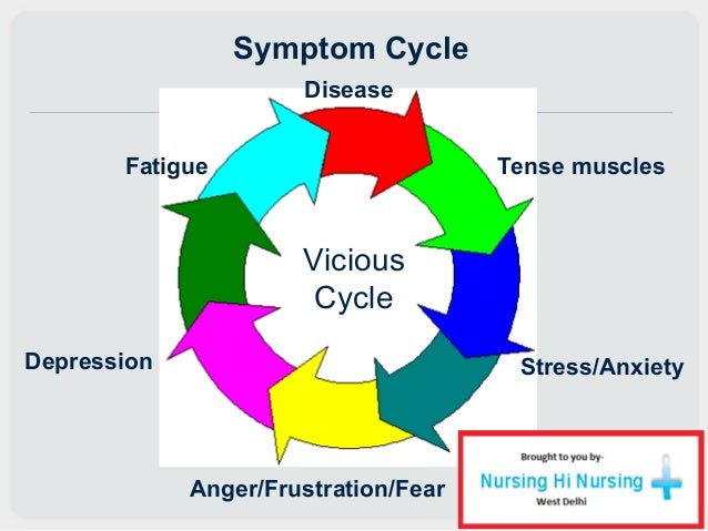
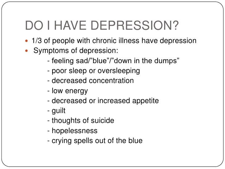
I want the words, "mental illness" to not hold a stigma, but they do. We have been taught to stay away from these words lest they conjure up a padded room, where we are fed bowls of oatmeal, under a locked door, in a straight jacket. However, there are "mental illnesses" out there that can be treated so that we do not have to go to some place like that. While the mental healthcare in our country isn't the best, it's come a long way. I suffer from anxiety and depression but I go to a psychiatrist in order to treat it. I have to take care of my mind just like I try and take care of my body, even though Gastroparesis makes it harder, like any chronic illness would.

NOTE: THIS BLOG DOES NOT SUBSTITUTE FOR A DOCTOR. PLEASE, IF YOU ARE HAVING ANY DEPRESSION, ANXIETY, AND OTHER MENTAL HEALTH CONCERNS, PLEASE SEE YOUR DOCTOR ASAP! THIS IS SERIOUS AND IT'S GOOD TO HAVE AN OUTSIDE OF THE FAMILY, WHOM IS UNBIASED, AND WILL HELP YOU. AGAIN, WE ARE NOT DOCTORS SO PLEASE CHECK WITH THEM FOR ANY MEDICAL QUESTIONS YOU MAY HAVE OR IDEAS YOU MAY HAVE FROM THIS BLOG.

When you have a chronic illness, it can be isolating, depressing, and cause anxiety. I wrote about this in January 2017, and here is Part One of this article if you would like to read it before you delve into this one:

http://www.emilysstomach.com/2017/01/mental-health-and-gastroparesis-part-1.html


Image Source: HERE


Anxiety and/or Depression with A Chronic Illness

I apologize for taking an entire year to write a part two to this article. I, myself, have had a rough year like everyone else seemed to in the GP Community. I lost A LOT of friends last year in my support groups, people I started support groups with, and people I talked to regularly. I logged into Facebook this morning to check on my groups, as I've been sick with pneumonia on top of Gastroparesis for the past week and a half (before that, my husband and I were passing a respiratory virus back and forth) and found another one of my friends had died. She was someone I've known since I first started advocating for Gastroparesis. She was apart of the handful of women who helped start Gastroparesis support groups and build up the Gastroparesis Community. I wanted to dedicated this article to her, and the other friends I've lost. Last year, we lost 48 people.


Image Source: HERE


This year, so far, I think the total is eight people. It's heartbreaking. We need better treatments. Having invasive "treatments" like feeding tubes, which can cause infections and worse, and then on the other end of the spectrum, medicine like Reglan, which can lead to irreversible, neurological disorders. We need medication to help our stomachs, to keep them from cramping so badly, to help them contract. Most of all, we need doctors to not only acknowledge our pain, but the depression and anxiety that comes with having a chronic illness. So, I am going to focus on mental health and how to get help


Image Source:CDC


I have noticed, and it seems like more and more lately, that people are comparing chronic illness to other chronic illnesses. This is something that I never really understood. I mean, if you have a chronic illness and I have a chronic illness, why compare them? Don't we both have chronic illnesses? Why does one have to be worse than the other one when they are both miserable in similar ways? No, they may not be the exact same illnesses, but we have a lot in common. We should lift each other up and support one another, not diminish each other's plights. Plus, the stress of having people tell you that your illness is not as bad as other illnesses, which seems worse to me if you are told that by a family member and/or support groups, and that hurts.

For example, a friend of someone in my family wrote on my Facebook post (I was upset because I had been throwing up all day), "At least you don't have cancer." No, I do not have cancer but I have watched my family members and good friends die from cancer. That was a horrible thing to say! Gastroparesis may not be cancer, but complications of it have killed my friends. It just makes people, like us, feel worse and that can set off flares of our Gastroparesis, but I call them, "Attacks," because it feels like my body is waging war on itself.


Image Source: On the Image


That made me feel so much worse, physically, but definitely worse mentally. I see a psychiatrist, and I've been seeing him since 2004. I do this because mental health is important too, not just physical health. I want to make sure I can talk to someone and to get the help I need. I have severe anxiety and really bad social anxiety. That stems from my Gastroparesis. Most of my friends are sympathetic vomiters, and I vomit A LOT, so I'm scared to go to social functions, and scared I might cause a vomiting conga line.


The Brain in Your Gut (90% of serotonin in made here): http://www.emilysstomach.com/2017/09/the-brain-in-your-gut.html

Suicide and Chronic Illness: http://www.emilysstomach.com/2014/10/sucide-and-chronic-illness.html

When Telling Someone They Look Great Becomes an Insult: http://www.emilysstomach.com/2013/01/when-telling-someone-they-look-great.html


Image Source: HERE


Staying Positive When You are Stressed and/Depressed

**NOTE I am not a doctor. If you are depressed and/or suffer from anxiety, please talk to your doctor. They can help you further. This article is just suggestions and advice if you are going through a difficult situation, it does not get rid of or substitute for a doctor's care and advice**

I have written on this subject in different ways. Here are articles I have written in the past that I wanted to share before I start this article:

SUICIDE AND CHRONIC ILLNESS.

THE GRIEVING PROCESS FOR A CHRONIC ILLNESS AND HOW TO OVERCOME IT.

HOW TO STAY POSITIVE, ESPECIALLY IN DIFFICULT SITUATIONS.

LOSING A LOVED ONE TO CHRONIC ILLNESS AND HANDLING GRIEF.

SLEEP DEPRIVATION'S EFFECTS ON THE BRAIN.

FEELING GOOD EVEN WHEN YOU'RE FEELING DOWN.

SEVERE EMOTIONAL AND PHYSICAL PAIN.

ISOLATION AND SADNESS.


Source Image: HERE


Chronic Illness and Depression

**AUTHOR'S NOTE:** First and Foremost, depression isn't as simple as just be happy. It runs deep and it's hard to stay positive or enjoy the things you love. I've been struggling with depression for the past year. I find that I'm having more and more trouble sleeping at night, so not only am I depressed, but sleep deprived and exhausted. That will definitely NOT help a mental state in any way. As a matter of fact, it makes depression worse.


I have said things to friends on Facebook that might have been misconstrued because it's hard to convey tone over the Internet and also, because I may have worded it wrong (again, not trying to make excuses, I own up to when I make a mistake) because vomiting for the past forty-eight hours with no sleep, tends to effect the way you word things. Additionally, being chronically ill on top of that, plus a death in the family, not to mention another death five months ago, plus cyber bullies, mean comments on posts, and me being scared to log into Facebook because I don't know what the next horrible thing is going to be. This has really taken it's toll on me.


I know that I need to call my doctor tomorrow, and I will, but until then, I wanted to take all of this negativity and use it to create something positive. I'm human, and I make mistakes (probably more so lately because everything just happened at once, and it's A LOT to handle) and I am not perfect. I ask that you be patient with me, as I am trying to do my best.


I am so sorry to those of you I may have offended, and I hope in your heart, you can forgive me. I did not mean to come across as mean, as uncaring, and that I care about group numbers, page likes, or blog views than the GPers themselves; That is NOT true at all! Far from it! All I have ever wanted to do was spread awareness and help people. If I could just help one person, then all of this was worth it. I don't want people to think I value numbers over their well being.


I've never been that type of person and I don't want to come across that way. I deeply care about the GP Community and do my best to contribute with positive, educational, and articles, to try and contribute a little bit to the community to let people know they're not alone. The strongest words I can offer are "I believe you." If I can touch one person to help them or make them laugh, make them forget about the awfulness for a moment, then I feel like I have helped some. I'm just really depressed and down lately because I feel like the GP Community does not want me involved with them. Furthermore, I have panic attacks when I go to log in now, because like I said previously, I don't know what awful thing I'll have to face when I do log in. I'm exhausted, stressed, and have cried more in the past few months than I have in the past two years. I just feel lost and alone. The isolation is also not good for me but I don't feel well enough to do anything since I'm recovering from the worst flare I've had since 2012 (not saying I've never dealt with bad flares before but this one has been worse than most). The last time I was this sick, I was hospitalized for eight days and diagnosed with Gastroparesis. I just feel helpless and hopeless. I feel like all of my positivity went out of the window in the past few months. Not only do I have to deal with bad things in my personal life, but on Facebook now as well. I'm just not sure what to do anymore. The drama that's on Facebook has constantly been linked to me. I've been the subject of it but I've never started drama for the sake of starting drama (why would I want to tear down a community I helped to build). Since I am the subject of drama from different groups and different people, and the fact it keeps happening, makes people and organizations to take a step back and they tell me that I can't write for them anymore or do certain things because of all of the drama. That hurts more than anything. I can take attacks on me but they've started to effect the things I loved to do...and I'm not sure I can ever fix it. I will be honest, since it keeps happening, and I am the common denominator in all of it (you can check, I've never said a bad word publicly about anyone who has attacked me or the organizations who do not want any part of this mess. I can't blame them, really. Their organization comes first. But it's turned into a pattern now, so maybe it is me. I guess they want me off of Facebook. I just don't know how I can make this right. Maybe I can never make this right. What's done is done, and I feel like because of that, my happiness is in ashes and it feels like my soul has been crushed. I'm not over exaggerating, this is how I really feel. There has to be something wrong with me, because even though they're victim blaming, it's happening over and over again.


According to the World Journal of Gastroenterology,


Image Source: At Top


According to U.S. Pharmacist,

"A New Approach to Managing Gastroparesis

Manouchehr Saljoughian, PharmD, PhD
Department of Pharmacy
Alta Bates Summit Medical Center
Berkeley, California


US Pharm. 2019;44(2):32-34.


Gastroparesis is a chronic disorder that affects a significant subset of the population. Ordinarily, strong muscular contractions move food through the digestive tract. In gastroparesis, this mechanism is disrupted, and undigested food stays in the abdomen for a long time and makes a person feel nauseous with the urge to vomit. Gastroparesis can also cause a lack of appetite, which may lead to malnutrition, and patients who are not eating can expect discomfort, bloating, and heartburn.1

The pathophysiology behind gastroparesis is varied and depends on disease etiology. Vagal and/or autonomic neuropathy play an important role in the development of diabetic gastroparesis, and it is estimated to occur in up to 20% to 40% of patients with diabetes. Gastroparesis can cause problems with blood sugar levels and nutrition. Sometimes, it is a complication of diabetes, and some people may develop gastroparesis after surgery. Although there is no cure for gastroparesis, changes to the diet, along with medication, can offer some relief.1,2

Certain medications, such as some antidepressants, opioid pain relievers, and high blood pressure and allergy medications, can lead to slow gastric emptying and cause similar symptoms. For people who already have gastroparesis, these medications may make their condition worse. Women are more likely to develop gastroparesis than men, and it is reported that many people with gastroparesis do not have any noticeable signs or symptoms.1 In this article, we briefly review the symptoms, causes, complications, and management of gastroparesis.


Symptoms

Signs and symptoms of gastroparesis include a feeling of fullness after eating just a few bites, vomiting undigested food eaten a few hours earlier, acid reflux, abdominal bloating, abdominal pain, changes in blood sugar levels, lack of appetite, and weight loss.3
Causes and Risk Factors

There are several risk factors that are considered to play a role in the condition’s cause, such as vagus-nerve damage. The vagus nerve is the longest cranial nerve in the body and is responsible for many functions. It is especially essential for proper operation of the digestive tract. If the vagus nerve is damaged, transfer of food from the abdomen to the small intestine is reduced because the muscles will not operate properly.4

Type 1 and type 2 diabetes are known to damage the vagus nerve. Some autoimmune diseases and virus infections (e.g., HIV) are also believed to have a negative impact on the vagus nerve. In certain cases, the vagus nerve stops working properly due to drinking excessive alcohol. Surgical complications could also affect the vagus nerve.4

Other factors that can increase the risk of gastroparesis include abdominal or esophageal surgery, infection (usually a virus), certain medications that slow the rate of stomach emptying (such as narcotic pain medications), nervous system diseases (such as Parkinson’s disease or multiple sclerosis) and hypothyroidism.4 Complications resulting from gastroparesis are shown in TABLE 1.

Treatment of gastroparesis depends on the cause, the severity of symptoms and complications, and how well patients respond to different treatments. As a result, the main goals of treatment for gastroparesis are alleviation of symptoms, correction of malnutrition, and resumption of adequate oral intake of liquids and solids. Patients with severe nausea and vomiting might require hospitalization for IV fluid and electrolyte replacement, and IV-administered prokinetic and/or antiemetic drugs might be needed initially.5

Sometimes, treating the cause may stop the problem. If diabetes is causing gastroparesis, patients must control their blood glucose levels. Acute hyperglycemia may impair gastric motor function as well as inhibit the action of prokinetic drugs, such as erythromycin. In patients with type 1 diabetes, gastroparesis can be an indication for insulin-pump therapy.5

Most physicians recommend that patients have a low-fat and low-fiber diet, eat smaller portions frequently during the day, chew food properly, eat well-cooked food, avoid alcohol and carbonated water, and drink plenty of water.


Medication Therapy

Initial management of gastroparesis consists of dietary modification, optimization of glycemic control and hydration, and in patients with continued symptoms, pharmacologic therapy with prokinetics and antiemetics.

Metoclopramide: This first-line therapy for gastroparesis is a dopamine 2 receptor antagonist, a 5-HT4 agonist, and a weak 5-HT3 receptor antagonist. It improves gastric emptying by enhancing gastric antral contractions and decreasing postprandial fundus relaxation.6

Metoclopramide is also used short-term to treat heartburn caused by gastroesophageal reflux in people who have used other medications without symptom relief. Dosage is 10 mg to 15 mg orally up to four times a day, 30 minutes before each meal and at bedtime. Depending upon symptoms being treated and clinical response, dosage will be different. It is commonly used to treat and prevent nausea and vomiting.6

Erythromycin: This macrolide antibiotic has been available since the 1950s. It is rarely used as an antibiotic today and is primarily prescribed for its “prokinetic” effect on the gastrointestinal (GI) tract. It has been used successfully off-label for the treatment of gastroparesis and other GI hypomotility disorders. When erythromycin was used as an antibiotic, patients often complained that it caused abdominal pain. Researchers eventually determined that erythromycin stimulates motilin receptors in the GI tract. Motilin receptors stimulate GI contractions and result in increased GI motility. This medicine also increases stomach-muscle contraction and may improve gastric emptying.7

Both oral and IV erythromycin have been used for its prokinetic effect. The IV form is generally reserved for acute conditions. The oral form is usually given in lower dosages than required for antibiotic effects (i.e., 150 mg-250 mg orally 3 to 4 times a day given 30 minutes before a meal). The oral form has been shown to work rapidly and can be substituted when the IV form is unavailable.7

Domperidone: This medication is used to treat nausea and vomiting as well as complaints of the stomach that occur with delayed emptying. It is used in patients whose symptoms fail to respond to metoclopramide or with side effects to metoclopramide. Domperidone is a dopamine 2 antagonist and is available for use only under a special program administered by the FDA. Each film-coated tablet contains 10-mg domperidone base. It should be taken 15 to 30 minutes before meals and, if necessary, before sleep. If taken after meals, absorption is somewhat delayed. Domperidone is taken by adults and adolescents aged 12 years or older.8

Cisapride: This 5-HT4 agonist stimulates antral and duodenal motility and accelerates gastric emptying of solids and liquids, which, in open-label trials, has been maintained for up to 1 year. Although cisapride is better tolerated than metoclopramide, its use has been associated with important drug interactions with medications metabolized by the cytochrome P450-3A4 isoenzyme (e.g., macrolide antibiotics, antifungals, and phenothiazines), resulting in cardiac arrhythmias. In the United States, prescriptions for cisapride can only be filled through an investigational limited-access program from the manufacturer after providing documentation as to the patient’s need for cisapride and assessment of risk factors for cardiac arrhythmias (e.g., a QTc >450 ms).9

Antiemetics: Antiemetics are medicines that help relieve nausea and vomiting. Prescription antiemetics include ondansetron, prochlorperazine, and promethazine. Over-the-counter antiemetic medications include bismuth subsaliclate and diphenhydramine. Antiemetics do not improve gastric emptying. In addition, they have not been studied in the management of patients with gastroparesis, and their use in gastroparesis is based on their efficacy in controlling nonspecific nausea and vomiting and in chemotherapy-induced emesis. Diphenhydramine 12.5 mg to 25 mg is given orally or IV every 6 to 8 hours as needed and in patients with persistent symptoms. Ondansetron, a 5-HT3 antagonist, is given 4 mg to 8 mg orally three times daily. Prolongation of the QT interval and central side effects have limited the use of phenothiazines, such as prochlorperazine, to patients who remain symptomatic despite antihistamines and 5-HT3 antagonists.1,4,10

Tricyclic Antidepressants: Low-dose nortriptyline, a tricyclic antidepressant with low anticholinergic effects, has been demonstrated to decrease symptoms of nausea, vomiting, and abdominal pain in patients with diabetic and idiopathic gastroparesis. Certain antidepressants, such as mirtazapine, may help relieve nausea and vomiting. These medicines may not improve gastric emptying.11

Pain Medicines: Pain medicines that are not narcotic may reduce pain in the abdomen due to gastroparesis.

Gastric Electrical Stimulation: This procedure may be considered for compassionate treatment in patients with refractory symptoms, particularly nausea and vomiting with persisting symptoms despite antiemetic and prokinetic drug therapy for at least 1 year. Gastric electrical stimulation has been demonstrated to improve symptom severity and gastric emptying in patients with diabetes but not idiopathic or postsurgical gastroparesis. In the U.S., the gastric electrical neurostimulator has been approved as a humanitarian exemption device for diabetic and idiopathic gastroparesis.12


REFERENCES

1. Camilleri M, Parkman HP, Shafi MA, et al. Clinical guideline: management of gastroparesis. Am J Gastroenterol. 2013;108:18-37.
2. Wytiaz V, Homko C, Duffy F, et al. Foods provoking and alleviating symptoms in gastroparesis: patient experiences. Dig Dis Sci. 2015;60:1052-1058.
3. Homko CJ, Duffy F, Friedenberg FK, et al. Effect of dietary fat and food consistency on gastroparesis symptoms in patients with gastroparesis. Neurogastroenterol Motil. 2015;27:501-508.
4. Type 2 diabetes and gastroparesis. www.healthline.com/health/type-2-diabetes/gastroparesis. Accessed August 2018.
5. Parkman HP, Yates KP, Hasler WL, et al. Dietary intake and nutritional deficiencies in patients with diabetic or idiopathic gastroparesis. Gastroenterology. 2011;141:486-498.
6. Rao AS, Camilleri M. Review article: metoclopramide and tardive dyskinesia. Aliment Pharmacol Ther. 2010;31:11-19.
7. Maganti K, Onyemere K, Jones MP. Oral erythromycin and symptomatic relief of gastroparesis: a systematic review. Am J Gastroenterol. 2003;98:259-263.
8. Sugumar A, Singh A, Pasricha PJ. A systematic review of the efficacy of domperidone for the treatment of diabetic gastroparesis. Clin Gastroenterol Hepatol. 2008;6:726-733.
9. Abell TL, Camilleri M, DiMagno EP, et al. Long-term efficacy of oral cisapride in symptomatic upper gut dysmotility. Dig Dis Sci. 1991;36:616-620.
10. Youssef AS, Parkman HP, Nagar S. Drug-drug interactions in pharmacologic management of gastroparesis. Neurogastroenterol Motil. 2015;27:1528-1541.
11. Prakash C, Lustman PJ, Freedland KE, Clouse RE. Tricyclic antidepressants for functional nausea and vomiting: clinical outcome in 37 patients. Dig Dis Sci. 1998;43:1951-1956.
12. Heckert J, Sankineni A, Hughes WB, et al. Gastric electric stimulation for refractory gastroparesis: a prospective analysis of 151 patients at a single center. Dig Dis Sci. 2016;61:168-175.
To comment on this article, contact rdavidson@uspharmcist.co
Read More On: GASTROENTEROLOGY"

Gastroparesis Medical Studies Update; Join and/or Keep Up With Clinical Trials


Source: Unknown


Source: Unknown


Source: On The Image but one of my favorites on how to explain Gastroparesis.


Clinical Trials - We Need Better Treatments

I apologize for taking so long to write an article. I, myself, have had a rough year like everyone else seemed to in the GP Community. I lost A LOT of friends last year in my support groups, people I started support groups with, and people I talked to regularly. I logged into Facebook this morning to check on my groups, as I've been sick with pneumonia on top of Gastroparesis for the past week and a half (before that, my husband and I were passing a respiratory virus back and forth) and found another one of my friends had died. She was someone I've known since I first started advocating for Gastroparesis. She was apart of the handful of women who helped start Gastroparesis support groups and build up the Gastroparesis Community. I wanted to dedicated this article to her, and the other friends I've lost. Last year, we lost 48 people. This year, so far, I think the total is four or five people. It's heartbreaking. We need better treatments. Having invasive "treatments" like feeding tubes, which can cause infections and worse, and then on the other end of the spectrum, medicine like Reglan, which can lead to irreversible, neurological disorders. We need medication to help our stomachs, to keep them from cramping so badly, to help them contract.

We need SOME kind of help. I'm so upset at watching my friends suffer and die. I get scared every time I throw up, wondering if it's going to be the last time - if this is the time I'm going to rupture my esophagus and die. I get panic attacks logging on to Facebook, scared I might read another one of my friends has passed away, which always upsets me right away, not only because they passed, but I feel like when I take breaks from Facebook, that I'm letting them down for not being online. I feel like I didn't get to say what I wanted to say to them before they passed. Most of that is on me. I've been not terrified to log into Facebook, but just


If you are interested in joining a clinical trial for Gastroparesis, The National Institute of Diabetes, Digestive, and Kidney Diseases are having people sign up for one now: https://www.niddk.nih.gov/health-information/digestive-diseases/gastroparesis/clinical-trials


Centerwatch has a long list of clinical trials that need volunteers as well: https://www.centerwatch.com/clinical-trials/listings/condition/72/gastroparesis/


ClinicalTrials.gov has a current trial happening as we speak: https://clinicaltrials.gov/ct2/show/NCT03500354

The Trial Says,
"Brief Summary:

Gastroparesis is a chronic, morbid and costly neuromuscular disorder of the stomach characterized by delayed gastric emptying in the absence of gross structural abnormalities. The periprandial symptoms associated with this disease can preclude adequate oral intake and often lead to weight loss and nutritional deficiencies 1. These manifestations are largely due to impaired gastric accommodation of meals and delayed transfer of food boluses from the stomach into the duodenum2. Consequently, the investigators hypothesize that dietary supplementation with a low volume, hypercaloric nutritional drink can help prevent malnutrition, decrease symptom burden and improve health-related quality of life in this population. Due to the paucity of such a supplement, the investigators developed a novel nutritional drink designed to maximize tolerability in patients with gastroparesis . This nutritional drink was tested on healthy volunteers (phase I) and passed the palatability test. The investigators now aim to test the tolerability of this drink on gastroparesis patients.

Condition or disease Intervention/treatment Phase
Gastroparesis Dietary Supplement: Nutrient drink Not Applicable

Detailed Description:

Primary objective:

To evaluate the safety and tolerability of the nutritional drink in gastroparesis patients.

Secondary objective:

To evaluate the efficacy of the nutritional drink in gastroparesis patients.

Study Procedures This study will be a pilot, open-label, trial in gastroparesis patients. A total of 20 patients will be recruited from the gastroenterology gastroparesis clinic. If the volunteer meets eligibility criteria, a co-investigator will contact the patient to schedule a study visit with a nutritionist and obtain a written consent. The contact and screening information of patients that are successfully recruited will be documented, placed in the participant's study folder and stored in a locked cabinet in the research unit. Any information documented during the screening process for patients who do not meet basic eligibility criteria or do not wish to participate will be immediately destroyed.

Patients will be given enough supply of the nutrition drink for (4 weeks) and asked to consume 200 ml of the drink three times daily. A follow-up call will be scheduled on day 2, day 7 and at the end of the study to make sure patients are tolerating the drink. Participants will be allowed to consume water and food as desired during the study period but will need to maintain an accurate food diary for at least one week prior to enrollment and during the study (at 2 weeks and at 4 weeks) along with weight measurements at baseline, 2 weeks, 4 weeks and 6 weeks. The participants will be asked to complete a palatability questionnaire. They will also complete the Gastroparesis Cardinal Symptom Index (GCSI) daily diary and the PROMISE scale prior to enrollment as a baseline for their symptoms and again at 2 weeks, 4 weeks (end of the study) and 6 weeks. Changes in these scales from baseline will determine the efficacy and possibly side effects of the nutritional drink.

Study duration and number of study visits required of research participants:

4 weeks, initial study visit with a nutritionist for screening and consenting followed by 3 follow-up phone calls on day 2, day 7 and at 4 weeks (the end of the study) and a final study visit at 6 weeks (2 weeks after finishing the study)


Study Type : Interventional (Clinical Trial)
Estimated Enrollment : 20 participants
Intervention Model: Single Group Assignment
Intervention Model Description: Pilot feasibility open-label study
Masking: None (Open Label)
Primary Purpose: Treatment
Official Title: Nutritional Drink in Gastroparesis
Estimated Study Start Date : February 1, 2019
Estimated Primary Completion Date : May 2019
Estimated Study Completion Date : August 2019


Primary Outcome Measures:

Tolerability will be measured by the Palatability Questionnaire at 2 days [ Time Frame: 2 days post-intervention ]

Patients will be given enough supply of the nutrition drink for 4 weeks and asked to consume 200 ml of the drink three times daily. A follow-up call will be scheduled on day 2 of the study to make sure patients are tolerating the drink.

Tolerability will be measured by the Palatability Questionnaire which rates six items on a scale of 1 to 5 (1= strongly disagree and 5= strongly agree. The six items are:
The formula/supplement tasted very good.
The formula/supplement tasted very bad.
I had no problems drinking the supplement.
Drinking the supplement made me feel ill.
I could drink more of this supplement anytime
I would never drink more of this supplement again

Tolerability will be measured by the Palatability Questionnaire at 7 days [ Time Frame: 7 days post-intervention ]

Patients will be given enough supply of the nutrition drink for 4 weeks and asked to consume 200 ml of the drink three times daily. A follow-up call will be scheduled on day 7 of the study to make sure patients are tolerating the drink.

Tolerability will be measured by the Palatability Questionnaire which rates six items on a scale of 1 to 5 (1= strongly disagree and 5= strongly agree. The six items are:
The formula/supplement tasted very good.
The formula/supplement tasted very bad.
I had no problems drinking the supplement.
Drinking the supplement made me feel ill.
I could drink more of this supplement anytime
I would never drink more of this supplement again

Tolerability will be measured by the Palatability Questionnaire at 4 weeks [ Time Frame: 4 weeks post-intervention ]

Patients will be given enough supply of the nutrition drink for 4 weeks and asked to consume 200 ml of the drink three times daily. A follow-up call will be scheduled at 4 weeks to make sure patients are tolerating the drink.

Tolerability will be measured by the Palatability Questionnaire which rates six items on a scale of 1 to 5 (1= strongly disagree and 5= strongly agree. The six items are:
The formula/supplement tasted very good.
The formula/supplement tasted very bad.
I had no problems drinking the supplement.
Drinking the supplement made me feel ill.
I could drink more of this supplement anytime
I would never drink more of this supplement again

Safety will be measured by the NIH PROMISE scale at baseline [ Time Frame: Baseline ]

This will be measured by the NIH PROMISE scale. This is a 10 point scale (0=none and 10= most severe) that rates the following symptoms:
Pain, especially in the abdomen, chest or back
Abdominal distension (bloating, sensation of excess gas)
Difficulty eating, sensation of food being stuck in the stomach.
Difficulty with bowel movements (constipation or straining)
Nausea and/or vomiting
Thirst
Weakness, lack of energy, fatigue, difficulty moving.

Safety will be measured by the NIH PROMISE scale at 2 weeks [ Time Frame: 2 weeks post-intervention ]

This will be measured by the NIH PROMISE scale. This is a 10 point scale (0=none and 10= most severe) that rates the following symptoms:
Pain, especially in the abdomen, chest or back
Abdominal distension (bloating, sensation of excess gas)
Difficulty eating, sensation of food being stuck in the stomach.
Difficulty with bowel movements (constipation or straining)
Nausea and/or vomiting
Thirst
Weakness, lack of energy, fatigue, difficulty moving.

Safety will be measured by the NIH PROMISE scale at 4 weeks [ Time Frame: 4 weeks post-intervention ]

This will be measured by the NIH PROMISE scale. This is a 10 point scale (0=none and 10= most severe) that rates the following symptoms:
Pain, especially in the abdomen, chest or back
Abdominal distension (bloating, sensation of excess gas)
Difficulty eating, sensation of food being stuck in the stomach.
Difficulty with bowel movements (constipation or straining)
Nausea and/or vomiting
Thirst
Weakness, lack of energy, fatigue, difficulty moving.

Safety will be measured by the NIH PROMISE scale at 6 weeks [ Time Frame: 6 weeks post-intervention ]

This will be measured by the NIH PROMISE scale. This is a 10 point scale (0=none and 10= most severe) that rates the following symptoms:
Pain, especially in the abdomen, chest or back
Abdominal distension (bloating, sensation of excess gas)
Difficulty eating, sensation of food being stuck in the stomach.
Difficulty with bowel movements (constipation or straining)
Nausea and/or vomiting
Thirst
Weakness, lack of energy, fatigue, difficulty moving.


How Gastroparesis Acts in The Body. Source:https://www.pinterest.com/pin/278026976974184742/?lp=true


Secondary Outcome Measures:

Improvement in gastroparesis symptoms [ Time Frame: Baseline, 2, 4 and 6 weeks ]
Change in weight compared to baseline

Improvement in gastroparesis symptoms [ Time Frame: Baseline, 2, 4 and 6 weeks ]

Changes in the Gastroparesis Cardinal Symptom Index (GCSI) daily diary as compared to baseline. This is a six point severity scale (0-5 with 0= none and 5 = very severe) that rates the following symptoms
Nausea
Early satiety
Postprandial fullness
Bloating
Upper abdominal pain
Retching
Vomiting
Stomach fullness
Loss of appetite
Stomach or belly visibly large


Information from the National Library of Medicine

Choosing to participate in a study is an important personal decision. Talk with your doctor and family members or friends about deciding to join a study. To learn more about this study, you or your doctor may contact the study research staff using the contacts provided below. For general information, Learn About Clinical Studies.

Ages Eligible for Study: 16 Years and older (Child, Adult, Older Adult)
Sexes Eligible for Study: All
Gender Based Eligibility: Yes
Gender Eligibility Description: Female or male
Accepts Healthy Volunteers: No
Criteria

Inclusion Criteria:

Patients with gastroparesis confirmed with symptoms and a gastric emptying study.
Inability to maintain adequate caloric intake by standard dietary measures for gastroparesis due to gastrointestinal symptoms

Exclusion Criteria:

Recent diagnosis of disorder other than gastroparesis that could affect food intake
Oropharyngeal dysphagia or other condition with risk for aspiration from oral ingestion.
Allergic reactions to any of the ingredients of the nutritional drink
Current pregnancy. Pregnancy status will be determined by questioning the potential subject.
Patient with gastrostomy/jejunostomy tube feeds or on total parenteral nutrition
Currently taking any anti-coagulant


Information from the National Library of Medicine

To learn more about this study, you or your doctor may contact the study research staff using the contact information provided by the sponsor.

Please refer to this study by its ClinicalTrials.gov identifier (NCT number): NCT03500354

Contacts

Contact: Pankaj J Pasricha, MD 4105027173 ppasric1@jhmi.edu
Contact: Carmen Roberts 4105027173 ccroberts@jhmi.edu

Locations

United States, Maryland
Johns Hopkins University Active, not recruiting
Baltimore, Maryland, United States, 21287
Sponsors and Collaborators
Johns Hopkins University
Investigators

Principal Investigator: Pankaj J Pasricha, MD Johns Hopkins University
More Information
Go to


Responsible Party: Johns Hopkins University
ClinicalTrials.gov Identifier: NCT03500354 History of Changes
Other Study ID Numbers: IRB00157677
First Posted: April 17, 2018 Key Record Dates
Last Update Posted: December 19, 2018
Last Verified: December 2018
Individual Participant Data (IPD) Sharing Statement:
Plan to Share IPD: No


Studies a U.S. FDA-regulated Drug Product: No
Studies a U.S. FDA-regulated Device Product: No

Keywords provided by Johns Hopkins University:

malnutrition


Additional relevant MeSH terms:

Gastroparesis
Stomach Diseases
Gastrointestinal Diseases
Digestive System Diseases
Paralysis
Neurologic Manifestations
Signs and Symptoms"


Source:https://preferredresearchpartners.com/gastroparesis-infographic/

Sunday, December 30, 2018

No Evidence of Impaired Gastric Emptying in Early Huntington‘s Disease

According to the PLOS Currents Huntington,


"No Evidence of Impaired Gastric Emptying in Early Huntington‘s Disease
November 16, 2011 · Epidemiology


Authors

Carsten Saft
Jürgen Andrich
Marc Fälker
Sarah Gauda
Sina Küchler
Dirk Woitalla
Oliver Goetze


Abstract

Background: Several factors, such as dysphagia, an increased motor activity, increased metabolic rate and a hypermetabolic state have been discussed as contributing to weight loss even at the early stages of Huntington’s Disease (HD). Aim of this pilot study was to investigate gastric emptying as a possible reason for weight loss in HD.

Methods: 11 HD participants at early stages of the disease and matched controls were investigated by using the well-established and non-invasive 13C-octanoate breath test. The “Gastroparesis Cardinal Symptom Index” and the “Short-Form Leeds Dyspepsia Questionnaire” were used for clinical evaluation of gastroparesis or dyspepsia.

Results: When compared to standard values ​​given in literature and controls all HD patients had normal breath test results. There was no evidence of gastroparesis or dyspepsia. There was a correlation of breath test results with the cognitive and functional performance of HD participants.

Conclusion: According to our data, there is no evidence of impaired gastric emptying in early HD. We can not exclude that gastric emptying contributes to weight loss at more advanced stages of the disease.

Corresponding author: PD Dr. med. Carsten Saft, Department of Neurology, Huntington-Center NRW, St. Josef Hospital, Gudrunstrasse 56, 44791 Bochum, Germany, E-mail: carsten.saft@ruhr-uni-bochum.de

§ Carsten Saft and Jürgen Andrich contributed equally to this work
Funding Statement
The study was supported by a FoRUM grant, University of Bochum (AZ: F506-2006). Oliver Götze was supported by the DFG (Gö 13582/1).
Introduction

Weight loss is a main feature in Huntington’s disease (HD) and was found to be manifest even at early stages of the disease. [1][2][3][4][5] Multifactorial causes, such as decreased caloric intake due to dysphagia and a higher energy expenditure due to increased motor activity have been discussed as being a possible reason for weight loss especially at the advanced stages of the disease. [6][7][8][9][10] Using a whole body indirect calorimetry in both early stage HD patients and the R6/2 transgenic mouse model of HD, Goodman and colleagues were able to demonstrate that patients with early HD tended to have a negative energy balance for reasons not related to their movement disorder, which was paralleled in the transgenic R6/2 mice. [4] This leads to the assumption of an increased metabolic rate as a main reason for weight loss in HD, which is supported by other experiments in the transgenic R6/2 mice. [4][11][12] In a study investigating the direct relation between the number of CAG repeats in the mutant huntingtin gene and weight loss, Aziz and colleagues found a correlation between both of these factors and discussed a hypermetabolic state as being a reason for weight loss, occurring even at early stages of the disease. [13] They discussed a hypermetabolic state as being likely to stem directly from interference of the mutant protein with cellular energy homeostasis and thus reflecting fundamental pathologic mechanisms underlying HD and not to be secondary to hyperactivity. Since mutant Huntingtin (mtHtt) is not only expressed in the brain of HD patients, but also in the gastrointestinal (GI) tract, a recently published study investigated the GI tract in the R6/2 mice model for HD. This study describes a loss of enteric neuropepitdes, a decreased mucosal thickness and villius length and also an impaired gut motility, diarrhea, and malabsorption of food, suggesting that GI dysfunction plays an important role in weight loss in HD mice. [14]

In addition, gastrointestinal dysfunction is discussed as being the main reason for weight loss in Parkinson’s disease (PD). [15] In a study using a solid meal and the 13 C-sodium octanoate breath test for measurement of gastric emptying in patients with PD, Goetze and colleagues found 88% of PD patients suufered from delayed gastric emptying when compared with controls. The severity of motor impairment was associated with gastroparesis. [16] Several other studies confirm an impaired gastric emptying in PD, some of them with a rate of 100% of PD patients. [16][17][18][19][20][21] One study describes a 60% delay in gastric half emptying time in the PD patient group after a solid test meal using the non-invasive 13 C-sodium octanoate breath test for evaluation of gastric emptying. [17] Neuropathological findings suggest enteric dysfunction to be one of the initial pathophysiological events in PD. [16][22] Central and enteric nervous system involvement in PD is discussed as being a pathophysiologic basis for this dysfunction. [15]

Autonomic nervous dysfunction was found to be present in HD, too. [23] Thus, the aim of the current study was to investigate gastric emptying in early HD patients without medication as a possible additional reason for weight loss by using the well-established 13 C-octanoate breath test. [16][17][18][19][20][21][24]
Methods


Participants

11 manifest HD patients with genetically confirmed diagnosis and without any medication in at clinically early stages of the disease (Shoulson stage I/II) and 11 controls were recruited from the HD centre Bochum, Germany. [25] Participants with known concurrent gastrointestinal diseases or previous operations of the gastro-intestinal tract were excluded, as well as patients with other severe diseases, diabetes mellitus, severe respiratory dysfunction, and malignancies. Also participants with concurrent liver diseases or excessive alcohol consumption (50 g/d of ethanol) were excluded. All participants had lab parameters for ALT, AST, LDH, cholesterol and triglycerides within the normal range, as well as normal findings for the ultrasonography of the upper abdomen. Pregnant and breast-feeding women were excluded. All HD participants underwent neurological investigation and were scored according to the UHDRS items “motor scale” (MS), “total functional capacity” (TFC) “independence scale” (IS) and the items verbal fluency test, symbol digit test, interference test, color naming and color reading which were summarized as “cognitive score” (CS). [26] Fine motor skills were additionally measured by simple (tapping; higher motor impairment leads to lower test results) and complex (pegboard; higher motor impairment leads to higher test results) instrumental movement tests. [27][28][29][30] The severity of depressive symptoms was assessed by using the Beck’s depression inventory (BDI) and Hamilton depression rating scale. [31][32] Clinical characteristics of all HD patients are given in table 1. In addition we calculated the disease burden score (DBS = [CAG repeat – 35.5] x age) for each subject. [33] The study was approved by the ethic committee of the Ruhr-University Bochum, Germany (registration-number 2719). Participants gave informed written consent according to GCP/ICH.

Parameter HD Participants Controls
Age [yr] 42.4 ± 8.4 (29-57) 48.9 ± 9.6 (38-69)
Gender (male/female) 3/8 3/8
BMI 22.5 ± 3.5 (16-30) 26.5 ± 6.4 (19-42)
Weight [kg] 63.6 ± 14.1 (42-85) 84.5 ± 22.5 (54-128)
Height [cm] 166.8 ± 10.1 (153-183) 178.2 ± 9.4 (164-190)
AO motor 39 ± 8.6 (25-51) –
AO psychiatric 38 ± 20.9 (29-50)a –
CAG expanded 45 ± 2.9 (42-51) –
Disease burden score 386.59 ± 66.06 (273-483) –
Disease duration [yr] 4.2 ± 2.5 (0.1-9) –

UHDRS MS 30.8 ± 18.7 (5-72) –
UHDRS TFC 10.2 ± 1.9 (7-12) –
UHDRS IS 81.8 ± 9.8 (70-100) –
UHDRS CS 195.1 ± 79.0 (98-346) –
Verbal fluency 22.5 ± 18.2 (4-69) –
SDMT 27.2 ± 10.6 (16-44) –
Stroop color 47.1 ± 17.1 (26-74) –
Stroop word 68.1 ± 22.8 (32-100) –
Stroop interference 29.5 ± 16.1 (10-59) –
Hamilton 12.8 ± 10.0 (1-26) –
Beck depression inventory 12.3 ± 13.4 (0-39) –

Tapping dominant 129.2 ± 44.8 (47-198) –
Tapping non dominant 99.4 ± 33 (38-161) –
Pegboard dominant [sec] 68.6 ± 24.8 (42.2-120.9) –
Pegboard non dominant [sec] 80.9 ± 40.2 (43.9-184.0) –

Table 1: Clinical characteristics of 11 HD patients and 11 matched controls; values are given as mean ± SD; range (min-max) in brackets; Abbreviations: BMI – body mass index, yr – years, AO – age at onset, a n = 6; UHDRS – unified Huntington´s disease rating scale, MS – motor score TFC – total functional capacity, IS – independence scale, CS – cognitive sum score, SDMT – symbol digit modalities test; sec – seconds. * – significant differences.


Test meal and 13C-octanoate breath test technique

The 13 C-octanoate breath test was used in the same way as described earlier. [16][17][18][34] In summary: After an overnight fasting each participant received a solid test meal consisting of an egg omelet of one egg, 60 g of white bread, 5 g of margarine and 150 ml of water (14 g of proteins, 26 g of carbohydrates and 9 g of fat, 241 kcal) labeled with 100 mg of 13C-sodiumoctanoate (chemical purity of 99,7 % and an isotopic purity of 99,1 %) at 8 AM. Breath samples, which were expired in close aluminized plastic breath bags of 50 ml content were obtained before substrate administration at baseline and after 10, 20, 30, 45, 60, 75, 90, 105, 120, 135, 150, 165, 180, 200, 220 and 240 minutes. The subjects were kept in a relaxed sitting position during the octanoate breath test (OBT). Physical activity was restricted during the test. All subjects consumed their test meal within 10 minutes. The 13 C/ 12 C isotope ratio of the breath samples was analysed by isotope-selective nondispersive infrared spectrometer (NDIRS). The results were both expressed as delta (δ) value per mil (‰) and delta over baseline (dob = δ s – δ 0 ). Definition of the δ-value: δ s = (R S /R PDB -1) x 1000 [‰] with R s = 13 C/ 12 C isotope ratio in CO 2 in breath and R PDB = 0.0112372 = isotope ratio in reference (PDB = PeeDeeBelmnite, South Carolina; δ 0 = isotope ratio at baseline).


Mathematical analysis of 13CO2 excretion curves and statistical analysis

As regards the measuring of the proportion of the 13 C-sodium octanoate given by mouth that is metabolised the results were expressed as a percentage dose of 13 C recovered (PDR) over time for each time interval from which the cumulative PDR (cPDR), obtained by numerical integration from PDR values, was calculated for each time interval. This calculation is based on the formula as proposed by Ravussin. [35] CO 2 production rate was assumed as being 300 mmol per unit of body surface area per hour. The body surface area was calculated using the Haycock weight-height formula. [36] The evaluation of the OBT for gastric emptying was done by non-linear regression analysis of the 13 CO 2 -excretion curves (PDR) with the formula PDR(t) = at b e -ct . The expression ln a, as gastric emptying coefficient (GEC) is a reliable parameter to describe the rate at which the stomach empties. The percentage of 13 CO 2 cumulative values was fit using a model given by the formula cPDR(t) = m(1-e -kt ) ß , where y is cPDR at time t in hours and m, k and ß are regression estimated constants, with m being the total amount of 13 CO 2 when time is infinite. Half gastric emptying time (t 50 ) was calculated by taking PDR(t) equal to m/2 in the PDR equation which is expressed as t 50 = (-1/k)ln (1-2 -1/ß ). The Lag phase is expressed as t lag = 1/klnß. [37] Statistical analysis was carried out as a descriptive evaluation of GEC, t 50 (min), tl ag (min) and t peak (min) and characteristics of participants (mean ± SD).


Gastroparesis Cardinal Symptom Index (GCSI) and Short-Form Leeds Dyspepsia Questionnaire (SF-LDQ)

The well-validated Gastroparesis Cardinal Symptom Index (GCSI) was used for clinical evaluation of gastroparesis symptoms. GCSI quantifies nine symptoms in the three different subscales: nausea and vomiting, postprandial fullness, and bloating. [38] In addition patients were asked about the frequency and severity of their stomach complaints, heartburn, burping and nausea symptoms according to the Short-Form Leeds Dyspepsia Questionnaire (SF-LDQ). [39]

The data analysis and statistics were performed by using the commercial software program SPSS statistics 19. All measured parameters and clinical data were first analysed descriptively and they were when presented as mean ± SD. Normality of distribution of the data was tested with the one-sample Kolmogorov-Smirnov test. Data were analyzed using the independent t-test for comparison between HD participants and controls. Pearson correlation analysis was used for exploratory statistical calculations of the normal distributed data.
Results

As expected HD participants had a lower body mass index compared to controls. One HD patient had underweight with a body mass index of 16. There were however, no significant differences between groups concerning any of the clinical data (table 1). Breath test results and clinical data showed normal distribution except for gender.

Results of the 13 C-sodium octanoate breath test are given in table 2. 13 CO 2 -excretion curves (PDR) and the percentage of 13 CO 2 cumulative values (cPDR) showed normal excretion of 13 C. For PDR only PDR max for the maximum amount of 13 CO 2 -excretion reached during testing time is listed in table 2. There were no significant differences compared to controls for the values decisive for the evaluation of gastric emptying, such as PDR max , cPDR, GEC, t 50 (min) and tl ag (min; see table 2). Compared to standard values given in literature, the most important parameters t 50 and tl ag were within normal range (t 50 < 200 min and tl ag <130 min; no data is available in literature for GEC, cPDR and PDR max ) and none of the patients had abnormal breath test results (see figure 1). [24] OBT Parameter Results HD Results Controls PDR max 9.76 ± 2.866.32 – 14.18 9.85 ± 2.615.58 – 14.09 cPDR 23.64 ± 7.92(14.72 – 37.0) 25.38 ± 7.92(15.51 – 34.94) GEC 2.96 ± 0.84(0.95 – 3.77) 2.89 ± 0.36(2.42 – 3.27) t 50 129.26 ± 38.84(77.15 – 197.60) 135.88 ± 22.27(95.74 – 167.47) t lag 85.45 ± 25.14(55.95 – 123.42) 80.74 ± 17.33(56.31 – 109.05) Table 2: 13 C-sodium octanoate breath test results; values are given as mean ± SD; range (min-max) in brackets, Abbreviations: PDR max for the maximum amount of 13 CO 2 -excretion reached during testing time [%]; cPDR – cumulative exhaled 13 CO 2 (cPDR [%]) after 240 minutes; GEC – gastric emptying coefficient; t peak – time to highest exhaled 13 CO 2 value [min]; t 50 – half gastric emptying time [min]; t lag – Lag phase [min]. * – significant differences. Fig. 1: Gastric emptying of solids measured by 13 C sodium octanoate breath test presented as individual lag phase (tl ag ) and gastric half emptying time (t50) in 11 HD participants (controls not shown). The normal t 50 range reported from literature (<200 min) is shown by the dotted line. A normal t lag range is reported to be below 130 min. [24] Gastroparesis Cardinal Symptom Index (GCSI) was 0.3855 (SEM ± 0.48; range 0 – 1.28) and Short-Form Leeds Dyspepsia Questionnaire (SF-LDQ) was 0.8182 (SEM ± 1.83; range 0 – 6) for HD participants. Thus, both questionnaire results were in line with published data from healthy controls, without clinical evidence of gastroparesis or dyspepsia. [38][39] GCSI was 0.3027 (SEM ± 0.30; range 0 – 0.83) and SF-LDQ was 2.273 (SEM ± 2.195; range 0 – 7) for controls. Differences were not significant (data not shown). Explorative correlation analysis of breath test results given in table 2 with clinical symptoms from table 1 showed no significant correlation, except for the cognitive sum score and t 50 (p 0.018, r -.692) and tl ag (p 0.019, r -.688), as well as for PDR max and the total functional capacity (TFC; p 0.014, r .712; no analysis of the cognitive subtests was done; see figure 2). Especially no correlation to motor symptoms was found. Fig. 2: A strong relation can be seen between gastric emptying of solids measured by 13 C sodium octanoate breath test A strong relation can be seen between gastric emptying of solids measured by 13 C sodium octanoate breath test, presented as individual t lag (a) and t 50 (b) in minutes and the cognitive sum score of the UHDRS (t 50 – p 0.018; r – .692 and t lag – p 0.019; r – .688), as well as for PDR max (c) and total functional capacity (TFC; p 0.014, r .712). The correlation analysis of GCSI and SF-LDQ with clinical symptoms showed no significant correlation with any of the clinical characteristics from table 1. Discussion Several factors such as dysphagia, an increased motor activity, an increased metabolic rate and hypermetabolic state have been discussed as contributing to weight loss even at early stages of HD. In addition, a recently published study also suggested gastrointestinal tract dysfunction as a reason for weight loss in a Huntington mouse model, similar to findings for Parkinson’s disease (PD). [14][15][21] Several studies describe a delay in gastric emptying for 88% or even for up to 100% of PD patients. [16][17][18][19][20][21] Contrary to this, in our pilot study on HD patients did not provide any evidence of impaired gastric emptying by using a solid meal and the 13 C-sodium octanoate breath test. There were no significant differences compared to controls and also compared to standard values given in literature all parameters were within normal range. In addition, we had no clinical evidence of gastroparesis or dyspepsia symptoms by using the “Gastroparesis Cardinal Symptom Index” and “Short-Form Leeds Dyspepsia Questionnaire” in our cohort. Thus, our data contrast with data for PD, but also with data for HD mice. As a possible explanation, gastric emptying may only contribute to weight loss more severe stages of HD. R6/2 HD mice usually show a very rapid course of the disease. A recent published study investigating the GI tract in a R6/2 mice model carrying a mean of 204 CAG-repeats describes several GI abnormalities, including an increased water content in R6/2 compared to feces in wild type mice from 8 weeks of age. The fecal output as a percentage of food intake however, was only significantly increased at 12 weeks, but not at 8 weeks. [14] This indicates that the occurrence of malabsorption of nutrients plays an important role in weight loss in HD mice only in the end stage. The study did not investigate early stages of the disease prior 8 weeks in the mice model. An earlier study from our group describes a high prevalence of gastritis or esophagitis as an accidental finding during PEG-placement, as a possible indication of gastrointestinal tract dysfunction in HD patients at advanced stages of the disease. [40] The findings in this study were also correlated with the duration and severity of the disease, also suggesting that gastrointestinal tract dysfunction might occur later in the course of the disease. We presumed that influences from the disease itself as well as secondary mechanisms like medication and general disability may contribute. [40] It was also the case in this study that the focus was not on early symptomatic patients. To summarize, the pilot data from our study suggest that impaired gastric emptying is not an early event in HD when compared to PD. We can not exclude that gastric emptying contributes to weight loss at more advanced stages of the disease. Surprisingly, we found a significant correlation for the cognitive sum score und the total functional capacity of the UHDRS and breath test results, such as t 50 (47.8% of variance), tl ag (47.4% of variance) and PDR max which usually shows the most precise quantification (50.6% of variance; see figure 2). This was not expected, since OBT results were within normal range. Cognitive decline, however, is a very early event in the course of HD. [41] In fact the cognitive sum score from our HD participants showed a broad range from 98-346 points with a mean of 195.1 points indicating a cognitive impairment in most of the patients. It is well known that the performance in UHDRS cognitive tests declined during disease progression, as did the functional capacity (TFC), which is highly dependent on cognitive tasks. [42][43] A decrement in mitochondrial function is discussed as contributing to age-dependent functional deficits in neurons and myocytes in normal aging and other neurological disorders, such as Alzheimer’s disease, accompanied with a cognitive decline. [44][45][46] Mitochondrial dysfunction is well known in HD and seems to be a relevant and early feature in the pathology. [47][48][49] Mutant htt (mtHtt) tends to aggregate in cytoplasm and nucleus of neurons as well as non-neuronal tissues including the liver. [50][51][52][53][54][55] Within the mitochondria, octanoic acid undergoes b-oxidation. Octanic acid generates acetyl coenzyme A which enters the Krebs cycle and is oxidized to CO 2 . Therefore breath tests based on octanoate, usually used to assess gastric emptying, should also reflect mitochondrial function. [56] Thus, one can speculate that a correlation of OBT results with results of cognitive tasks might reflect a parallel decline in cognitive and mitochondrial function. To our knowledge this is the first study dealing with gastrointestinal track dysfunction in HD in vivo. A limitation of our study is the relative small number of participants. To exclude drug effects we only included patients without any medication and without serious comorbidities. On the other hand, due to the fact that this is a very rare group of patients it is a strength of our study that we can exclude medication effects. Competing interests The authors declare that they have no competing interests. Ethics The local ethics committee of the university approved this study. Acknowledgements We are grateful to all patients for participation. References Djousse L, Knowlton B, Cupples LA, Marder K, Shoulson I, Myers RH. Weight loss in early stage of Huntington's disease. Neurology 2002;59:1325-1330. Sanberg PR, Fibiger HC, Mark RF. Body weight and dietary factors in Huntington's disease patients compared with matched controls. Med J Aust 1981;1:407-409. Trejo A, Tarrats RM, Alonso ME, Boll MC, Ochoa A, Velasquez L. Assessment of the nutrition status of patients with Huntington's disease. Nutrition 2004;20:192-196. Goodman AO, Murgatroyd PR, Medina-Gomez G, et al. The metabolic profile of early Huntington's disease--a combined human and transgenic mouse study. Exp Neurol 2008;210:691-698. Mochel F, Charles P, Seguin F, et al. Early energy deficit in Huntington disease: identification of a plasma biomarker traceable during disease progression. PLoS One 2007;2:e647. Hamilton JM, Wolfson T, Peavy GM, Jacobson MW, Corey-Bloom J. Rate and correlates of weight change in Huntington's disease. J Neurol Neurosurg Psychiatry 2004;75:209-212. Nance MA, Sanders G. Characteristics of individuals with Huntington disease in long-term care. Mov Disord 1996;11:542-548. Gaba AM, Zhang K, Marder K, Moskowitz CB, Werner P, Boozer CN. Energy balance in early-stage Huntington disease. Am J Clin Nutr 2005;81:1335-1341. Mahant N, McCusker EA, Byth K, Graham S. Huntington's disease: clinical correlates of disability and progression. Neurology 2003;61:1085-1092. Pratley RE, Salbe AD, Ravussin E, Caviness JN. Higher sedentary energy expenditure in patients with Huntington's disease. Ann Neurol 2000;47:64-70. van der Burg JM, Bacos K, Wood NI, et al. Increased metabolism in the R6/2 mouse model of Huntington's disease. Neurobiol Dis 2008;29:41-51. Weydt P, Pineda VV, Torrence AE, et al. Thermoregulatory and metabolic defects in Huntington's disease transgenic mice implicate PGC-1alpha in Huntington's disease neurodegeneration. Cell Metab 2006;4:349-362. Aziz NA, van der Burg JM, Landwehrmeyer GB, Brundin P, Stijnen T, Roos RA. Weight loss in Huntington disease increases with higher CAG repeat number. Neurology 2008;71:1506-1513. van der Burg JM, Winqvist A, Aziz NA, et al. Gastrointestinal dysfunction contributes to weight loss in Huntington's disease mice. Neurobiol Dis;44:1-8. Pfeiffer RF. Gastrointestinal dysfunction in Parkinson's disease. Parkinsonism Relat Disord;17:10-15. Goetze O, Nikodem AB, Wiezcorek J, et al. Predictors of gastric emptying in Parkinson's disease. Neurogastroenterol Motil 2006;18:369-375. Goetze O, Wieczorek J, Mueller T, Przuntek H, Schmidt WE, Woitalla D. Impaired gastric emptying of a solid test meal in patients with Parkinson's disease using 13C-sodium octanoate breath test. Neurosci Lett 2005;375:170-173. Muller T, Erdmann C, Bremen D, et al. Impact of gastric emptying on levodopa pharmacokinetics in Parkinson disease patients. Clin Neuropharmacol 2006;29:61-67. Woitalla D, Goetze O. Treatment approaches of gastrointestinal dysfunction in Parkinson's disease, therapeutical options and future perspectives. J Neurol Sci. Djaldetti R, Baron J, Ziv I, Melamed E. Gastric emptying in Parkinson's disease: patients with and without response fluctuations. Neurology 1996;46:1051-1054. Pfeiffer RF. Gastrointestinal dysfunction in Parkinson's disease. Lancet Neurol 2003;2:107-116. Braak H, Del Tredici K, Rub U, de Vos RA, Jansen Steur EN, Braak E. Staging of brain pathology related to sporadic Parkinson's disease. Neurobiol Aging 2003;24:197-211. Andrich J, Schmitz T, Saft C, et al. Autonomic nervous system function in Huntington's disease. J Neurol Neurosurg Psychiatry 2002;72:726-731. Keller J, Franke A, Storr M, Wiedbrauck F, Schirra J. [Clinically relevant breath tests in gastroenterological diagnostics--recommendations of the German Society for Neurogastroenterology and Motility as well as the German Society for Digestive and Metabolic Diseases]. Z Gastroenterol 2005;43:1071-1090. Shoulson I. Huntington disease: functional capacities in patients treated with neuroleptic and antidepressant drugs. Neurology 1981;31:1333-1335. Huntington Study Group. Unified Huntington's Disease Rating Scale: reliability and consistency. Mov Disord 1996;11:136-142. Andrich J, Saft C, Ostholt N, Muller T. Assessment of simple movements and progression of Huntington's disease. J Neurol Neurosurg Psychiatry 2007;78:405-407. Andrich J, Saft C, Ostholt N, Muller T. Complex movement behaviour and progression of Huntington's disease. Neurosci Lett 2007;416:272-274. Saft C, Andrich J, Meisel NM, Przuntek H, Muller T. Assessment of simple movements reflects impairment in Huntington's disease. Mov Disord 2006;21:1208-1212. Saft C, Andrich J, Meisel NM, Przuntek H, Muller T. Assessment of complex movements reflects dysfunction in Huntington's disease. J Neurol 2003;250:1469-1474. Hamilton M. A rating scale for depression. J Neurol Neurosurg Psychiatry 1960;23:56-62. Beck AT, Ward CH, Mendelson M, Mock J, Erbaugh J. An inventory for measuring depression. Arch Gen Psychiatry 1961;4:561-571. Penney JB, Jr., Vonsattel JP, MacDonald ME, Gusella JF, Myers RH. CAG repeat number governs the development rate of pathology in Huntington's disease. Ann Neurol 1997;41:689-692. Muller T, Woitalla D, Goetze O, Erdmann C. Entacapone improves absorption of a coadministered salt in patients with Parkinson's disease. Mov Disord 2008;23:1458-1461. Ravussin E, Pahud P, Thelin-Doerner A, Arnaud MJ, Jequier E. Substrate utilization during prolonged exercise after ingestion of 13C-glucose in obese and control subjects. Int J Obes 1980;4:235-242. Haycock GB, Schwartz GJ, Wisotsky DH. Geometric method for measuring body surface area: a height-weight formula validated in infants, children, and adults. J Pediatr 1978;93:62-66. Ghoos YF, Maes BD, Geypens BJ, et al. Measurement of gastric emptying rate of solids by means of a carbon-labeled octanoic acid breath test. Gastroenterology 1993;104:1640-1647. Revicki DA, Rentz AM, Dubois D, et al. Development and validation of a patient-assessed gastroparesis symptom severity measure: the Gastroparesis Cardinal Symptom Index. Aliment Pharmacol Ther 2003;18:141-150. Fraser A, Delaney BC, Ford AC, Qume M, Moayyedi P. The Short-Form Leeds Dyspepsia Questionnaire validation study. Aliment Pharmacol Ther 2007;25:477-486. Andrich JE, Wobben M, Klotz P, Goetze O, Saft C. Upper gastrointestinal findings in Huntington's disease: patients suffer but do not complain. J Neural Transm 2009;116:1607-1611. Stout JC, Paulsen JS, Queller S, et al. Neurocognitive signs in prodromal Huntington disease. Neuropsychology;25:1-14. Beglinger LJ, Duff K, Allison J, et al. Cognitive change in patients with Huntington disease on the Repeatable Battery for the Assessment of Neuropsychological Status. J Clin Exp Neuropsychol 2010;32:573-578. Beglinger LJ, O'Rourke JJ, Wang C, Langbehn DR, Duff K, Paulsen JS. Earliest functional declines in Huntington disease. Psychiatry Res;178:414-418. Bishop NA, Lu T, Yankner BA. Neural mechanisms of ageing and cognitive decline. Nature 2010;464:529-535. Glade MJ. Oxidative stress and cognitive longevity. Nutrition 2010;26:595-603. Sultana R, Butterfield DA. Oxidatively modified, mitochondria-relevant brain proteins in subjects with Alzheimer disease and mild cognitive impairment. J Bioenerg Biomembr 2009;41:441-446. Bossy-Wetzel E, Petrilli A, Knott AB. Mutant huntingtin and mitochondrial dysfunction. Trends Neurosci 2008;31:609-616. Saft C, Zange J, Andrich J, et al. Mitochondrial impairment in patients and asymptomatic mutation carriers of Huntington's disease. Mov Disord 2005;20:674-679. Taherzadeh-Fard E, Saft C, Akkad DA, et al. PGC-1alpha downstream transcription factors NRF-1 and TFAM are genetic modifiers of Huntington disease. Mol Neurodegener;6:32. Sassone J, Colciago C, Cislaghi G, Silani V, Ciammola A. Huntington's disease: the current state of research with peripheral tissues. Exp Neurol 2009;219:385-397. Browne SE. Mitochondria and Huntington's disease pathogenesis: insight from genetic and chemical models. Ann N Y Acad Sci 2008;1147:358-382. Davies SW, Turmaine M, Cozens BA, et al. Formation of neuronal intranuclear inclusions underlies the neurological dysfunction in mice transgenic for the HD mutation. Cell 1997;90:537-548. Sathasivam K, Hobbs C, Turmaine M, et al. Formation of polyglutamine inclusions in non-CNS tissue. Hum Mol Genet 1999;8:813-822. Scherzinger E, Lurz R, Turmaine M, et al. Huntingtin-encoded polyglutamine expansions form amyloid-like protein aggregates in vitro and in vivo. Cell 1997;90:549-558. Squitieri F, Cannella M, Sgarbi G, et al. Severe ultrastructural mitochondrial changes in lymphoblasts homozygous for Huntington disease mutation. Mech Ageing Dev 2006;127:217-220. Grattagliano I, Lauterburg BH, Palasciano G, Portincasa P. 13C-breath tests for clinical investigation of liver mitochondrial function. Eur J Clin Invest;40:843-850."

Tuesday, December 18, 2018

Myasthenia Gravis


What is Myasthenia Gravis?

Myasthenia gravis is a chronic autoimmune neuromuscular disease that causes weakness in the skeletal muscles, which are responsible for breathing and moving parts of the body, including the arms and legs. The name myasthenia gravis, which is Latin and Greek in origin, means 'grave, or serious, muscle weakness.'

The hallmark of myasthenia gravis is muscle weakness that worsens after periods of activity and improves after periods of rest. Certain muscles such as those that control eye and eyelid movement, facial expression, chewing, talking, and swallowing are often (but not always) involved in the disorder. The muscles that control breathing and neck and limb movements may also be affected.

There is no known cure but with current therapies most cases of myasthenia gravis are not as 'grave' as the name implies. Available treatments can control symptoms and often allow people to have a relatively high quality of life. Most individuals with the condition have a normal life expectancy.


What Causes Myasthenia Gravis?

Myasthenia gravis is caused by an error in the transmission of nerve impulses to muscles. It occurs when normal communication between the nerve and muscle is interrupted at the neuromuscular junction—the place where nerve cells connect with the muscles they control.

Neurotransmitters are chemicals that neurons, or brain cells, use to communicate information. Normally when electrical signals or impulses travel down a motor nerve, the nerve endings release a neurotransmitter called acetylcholine. Acetylcholine travels from the nerve ending and binds to acetylcholine receptors on the muscle. The binding of acetylcholine to its receptor activates the muscle and causes a muscle contraction.

In myasthenia gravis, antibodies (immune proteins) block, alter, or destroy the receptors for acetylcholine at the neuromuscular junction, which prevents the muscle from contracting. In most individuals with myasthenia gravis, this is caused by antibodies to the acetylcholine receptor itself. However, antibodies to other proteins, such as MuSK (Muscle-Specific Kinase) protein, can also lead to impaired transmission at the neuromuscular junction.

These antibodies are produced by the body's own immune system. Myasthenia gravis is an autoimmune disease because the immune system—which normally protects the body from foreign organisms—mistakenly attacks itself.

The thymus is a gland that controls immune function and maybe associated with myasthenia gravis. Located in the chest behind the breast bone, the gland is largest in children. It grows gradually until puberty, and then gets smaller and is replaced by fat. Throughout childhood, the thymus plays an important role in the development of the immune system because it is responsible for producing T-lymphocytes or T cells, a specific type of white blood cell that protects the body from viruses and infections.

In many adults with myasthenia gravis, the thymus gland remains large. People with the disease typically have clusters of immune cells in their thymus gland similar to lymphoid hyperplasia—a condition that usually only happens in the spleen and lymph nodes during an active immune response. Some individuals with myasthenia gravis develop thymomas (tumors of the thymus gland). Thymomas are most often harmless, but they can become cancerous.

The thymus gland plays a role in myasthenia gravis, but its function is not fully understood. Scientists believe that the thymus gland may give incorrect instructions to developing immune cells, ultimately causing the immune system to attack its own cells and tissues and produce acetylcholine receptor antibodies—setting the stage for the attack on neuromuscular transmission.


Source: On Image


What are the Symptoms of Myasthenia Gravis?

Although myasthenia gravis may affect any skeletal muscle, muscles that control eye and eyelid movement, facial expression, and swallowing are most frequently affected. The onset of the disorder may be sudden and symptoms often are not immediately recognized as myasthenia gravis.

In most cases, the first noticeable symptom is weakness of the eye muscles. In others, difficulty swallowing and slurred speech may be the first signs. The degree of muscle weakness involved in myasthenia gravis varies greatly among individuals, ranging from a localized form limited to eye muscles (ocular myasthenia), to a severe or generalized form in which many muscles—sometimes including those that control breathing—are affected.


Symptoms may include:

drooping of one or both eyelids (ptosis)
blurred or double vision (diplopia) due to weakness of the muscles that control eye movements
a change in facial expression
difficulty swallowing
shortness of breath
impaired speech (dysarthria)
weakness in the arms, hands, fingers, legs, and neck.


Who gets Myasthenia Gravis?

Myasthenia gravis affects both men and women and occurs across all racial and ethnic groups. It most commonly impacts young adult women (under 40) and older men (over 60), but it can occur at any age, including childhood. Myasthenia gravis is not inherited nor is it contagious. Occasionally, the disease may occur in more than one member of the same family.

Although myasthenia gravis is rarely seen in infants, the fetus may acquire antibodies from a mother affected with myasthenia gravis—a condition called neonatal myasthenia. Generally, neonatal myasthenia gravis is temporary and the child's symptoms usually disappear within two to three months after birth. Rarely, children of a healthy mother may develop congenital myasthenia. This is not an autoimmune disorder (it is caused by defective genes that produce abnormal proteins in the neuromuscular junction) and can cause similar symptoms to myasthenia gravis.


Source:HERE


How is Myasthenia Gravis Diagnosed?


A doctor may perform or order several tests to confirm the diagnosis, including:

A physical and neurological examination. A physician will first review an individual’s medical history and conduct a physical examination. In a neurological examination, the physician will check muscle strength and tone, coordination, sense of touch, and look for impairment of eye movements.

An EDROPHONIUM test. This test uses injections of edrophonium chloride to briefly relieve weakness in people with myasthenia gravis. The drug blocks the breakdown of acetylcholine and temporarily increases the levels of acetylcholine at the neuromuscular junction. It is usually used to test ocular muscle weakness.

A blood test. Most individuals with myasthenia gravis have abnormally elevated levels of acetylcholine receptor antibodies. A second antibody—called the anti-MuSK antibody—has been found in about half of individuals with myasthenia gravis who do not have acetylcholine receptor antibodies. A blood test can also detect this antibody. However, in some individuals with myasthenia gravis, neither of these antibodies is present. These individuals are said to have seronegative (negative antibody) myasthenia.

Electrodiagnostics. Diagnostic tests include repetitive nerve stimulation, which repeatedly stimulates a person’s nerves with small pulses of electricity to tire specific muscles. Muscle fibers in myasthenia gravis, as well as other neuromuscular disorders, do not respond as well to repeated electrical stimulation compared to muscles from normal individuals. Single fiber electromyography (EMG), considered the most sensitive test for myasthenia gravis, detects impaired nerve-to-muscle transmission. EMG can be very helpful in diagnosing mild cases of myasthenia gravis when other tests fail to demonstrate abnormalities.

Diagnostic imaging. Diagnostic imaging of the chest using computed tomography (CT) or magnetic resonance imaging (MRI) may identify the presence of a thymoma.


Source: HERE


Pulmonary function testing. Measuring breathing strength can help predict if respiration may fail and lead to a myasthenic crisis.
Because weakness is a common symptom of many other disorders, the diagnosis of myasthenia gravis is often missed or delayed (sometimes up to two years) in people who experience mild weakness or in those individuals whose weakness is restricted to only a few muscles.


Source: McGraw Hill


What is a Myasthenic Crisis?

A myasthenic crisis is a medical emergency that occurs when the muscles that control breathing weaken to the point where individuals require a ventilator to help them breathe.

Approximately 15 to 20 percent of people with myasthenia gravis experience at least one myasthenic crisis. This condition usually requires immediate medical attention and may be triggered by infection, stress, surgery, or an adverse reaction to medication. However, up to one-half of people may have no obvious cause for their myasthenic crisis. Certain medications have been shown to cause myasthenia gravis. However, sometimes these medications may still be used if it is more important to treat an underlying condition.


How is Myasthenia Gravis Treated?

Today, myasthenia gravis can generally be controlled. There are several therapies available to help reduce and improve muscle weakness.

THYMECTOMY. This operation to remove the thymus gland (which often is abnormal in individuals with myasthenia gravis) can reduce symptoms and may cure some people, possibly by rebalancing the immune system. A recent NINDS-funded study found that thymectomy is beneficial both for people with thymoma and those with no evidence of the tumors. The clinical trial followed 126 people with myasthenia gravis and no visible thymoma and found that the surgery reduced muscle weakness and the need for immunosuppressive drugs.

Anticholinesterase medications
. Medications to treat the disorder include anticholinesterase agents such as MESTINON or PYRIDOSTIGMINE, which slow the breakdown of acetylcholine at the neuromuscular junction and thereby improve neuromuscular transmission and increase muscle strength.

Immunosuppressive drugs. These drugs improve muscle strength by suppressing the production of abnormal antibodies. They include prednisone, azathioprine, mycophenolate mofetil, tacrolimus, and rituximab. The drugs can cause significant side effects and must be carefully monitored by a physician.

Plasmapheresis and intravenous immunoglobulin. These therapies may be options in severe cases of myasthenia gravis. Individuals can have antibodies in their plasma (a liquid component in blood) that attack the neuromuscular junction. These treatments remove the destructive antibodies, although their effectiveness usually only lasts for a few weeks to months.

Plasmapheresis is a procedure using a machine to remove harmful antibodies in plasma and replace them with good plasma or a plasma substitute.
Intravenous immunoglobulin is a highly concentrated injection of antibodies pooled from many healthy donors that temporarily changes the way the immune system operates. It works by binding to the antibodies that cause myasthenia gravis and removing them from circulation.


Source: On Image


What is the prognosis?

With treatment, most individuals with myasthenia can significantly improve their muscle weakness and lead normal or nearly normal lives.

Sometimes the severe weakness of myasthenia gravis may cause respiratory failure, which requires immediate emergency medical care.

Some cases of myasthenia gravis may go into remission—either temporarily or permanently—and muscle weakness may disappear completely so that medications can be discontinued. Stable, long-lasting complete remissions are the goal of thymectomy and may occur in about 50 percent of individuals who undergo this procedure.


What Research is Being Done?

The mission of the National Institute of Neurological Disorders and Stroke (NINDS) is to seek fundamental knowledge about the brain and nervous system and to use that knowledge to reduce the burden of neurological disease. The NINDS is a component of the National Institutes of Health (NIH), the leading supporter of biomedical research in the world.

Although there is no cure for myasthenia gravis, management of the disorder has improved over the past 30 years. There is a greater understanding about the structure and function of the neuromuscular junction, the fundamental aspects of the thymus gland and of autoimmunity, and the disorder itself. Technological advances have led to more timely and accurate diagnosis of myasthenia gravis and new and enhanced therapies have improved treatment options. Researchers are working to develop better medications, identify new ways to diagnose and treat individuals, and improve treatment options.


Medication

Some people with myasthenia gravis do not respond favorably to available treatment options, which usually include long-term suppression of the immune system. New drugs are being tested, either alone or in combination with existing drug therapies, to see if they are effective in treating the disease.

Studies are investigating the use of therapy targeting the B cells that make antibodies (rituximab) or the process by which acetylcholine antibodies injure the neuromuscular junction (eculizumab). The drugs have shown promise in initial clinical trials.


Diagnostics and biomarkers

In addition to developing new medications, researchers are trying to find better ways to diagnose and treat this disorder. For example, NINDS-funded researchers are exploring the assembly and function of connections between nerves and muscle fibers to understand the fundamental processes in neuromuscular development. This research could reveal new therapies for neuromuscular diseases like myasthenia gravis.

Researchers are also exploring better ways to treat myasthenia gravis by developing new tools to diagnose people with undetectable antibodies and identify potential biomarkers (signs that can help diagnose or measure the progression of a disease) to predict an individual’s response to immunosuppressive drugs.


Source: McGraw Hill


New treatment options

Findings from a recent NINDS-supported study yielded conclusive evidence about the benefits of surgery for individuals without thymoma, a subject that had been debated for decades. Researchers hope that this trial will become a model for rigorously testing other treatment options, and that other studies will continue to examine different therapies to see if they are superior to standard care options.


Where can I get more information?

For more information on neurological disorders or research programs funded by the National Institute of Neurological Disorders and Stroke, contact the Institute's Brain Resources and Information Network (BRAIN) at:

BRAIN
P.O. Box 5801
Bethesda, MD 20824
800-354-9424
www.ninds.nih.gov


More information about research on myasthenia gravis supported by NINDS and other NIH Institutes and Centers can be found using NIH RePORTER (projectreporter.nih.gov), a searchable database of current and past research projects supported by NIH and other federal agencies. RePORTER also includes links to publications and resources from these projects.


Information is also available from the following organizations:

Myasthenia Gravis Foundation of America, Inc.
355 Lexington Avenue, 15th Floor
New York, NY 10017
212-297-2156
800-541-5454
www.myasthenia.org

American Autoimmune Related Diseases Association
22100 Gratiot Avenue
Eastpointe, MI 48021
586-776-3900
www.aarda.org

Muscular Dystrophy Association
222 S. Riverside Plaza, Suite 1500
Chicago, IL 60606
800-572-1717
www.mda.org

U.S. National Library of Medicine
National Institutes of Health/DHHS
8600 Rockville Pike
Bethesda, MD 20894
301-594-5983
888-346-3656
www.nlm.nih.gov

NIH Publication No. 17-768

"Myasthenia Gravis Fact Sheet", NINDS, Publication date May 2017.

NIH Publication No. 17-768

Back to Myasthenia Gravis Information Page

See a list of all NINDS publications

Publicaciones en Español

Miastenia gravis

Prepared by:

Office of Communications and Public Liaison

National Institute of Neurological Disorders and Stroke

National Institutes of Health

Bethesda, MD 20892

NINDS health-related material is provided for information purposes only and does not necessarily represent endorsement by or an official position of the National Institute of Neurological Disorders and Stroke or any other Federal agency. Advice on the treatment or care of an individual patient should be obtained through consultation with a physician who has examined that patient or is familiar with that patient's medical history.

All NINDS-prepared information is in the public domain and may be freely copied. Credit to the NINDS or the NIH is appreciated.

PATIENT & CAREGIVER EDUCATION
Fact Sheets
Hope Through Research
Know Your Brain
Preventing Stroke
Understanding Sleep
The Life and Death of a Neuron
Genes At Work In The Brain
Order Publications
Date last modified: Fri, 2018-07-06 16:21"


Source:HERE


According to the CLEVELAND CLINIC,


"What is Myasthenia Gravis?

Myasthenia gravis (MG) is a neuromuscular disease, meaning that it affects the muscles and the nerves that control them. It is caused by a disorder in the immune system that causes the body to attack the area of the muscles where the nerves connect to them.

The immune system is the body’s natural defense against disease. Normally, when bacteria or other foreign substances enter the body, the immune system produces molecules called antibodies that attack the bacteria.

In people with myasthenia gravis, the immune system produces abnormal antibodies that prevent the muscles from receiving signals from the nerves that tell them when to relax or contract. This causes muscle weakness with symptoms that can include in double vision or blurred vision (eye muscle weakness), drooping eyelids (eyelid muscle weakness), difficulty with speaking and swallowing (throat muscle weakness) and weakness of the limbs.

When the immune system acts against healthy tissue by mistake, it is called an autoimmune disorder, with “auto” meaning “self. So myasthenia gravis is a neuromuscular autoimmune disease.

Myasthenia gravis is most common in young women and older men, but people of any age or either sex can get it.


Source:HERE


What Causes Myasthenia Gravis?

Scientists do not completely understand what triggers the autoimmune reaction in myasthenia gravis, but they do know that the thymus gland plays a role in the disease.

The thymus is a small gland that lies in the front part of the chest, beneath the breastbone, and extends into the lower part of the neck. It is most important early in life during immune system development.

A baby’s thymus gland weighs between .7 and 1.1 oz. The gland continues to grow and by puberty weighs 1.1 to 1.8 oz. The thymus gland’s job is thought to be completed by puberty, and after that it decreases in size. Over time, fat replaces portions of the gland. In older people the thymus weighs only .1 to .5 oz.

Tumors of the thymus gland are called thymomas (https://my.clevelandclinic.org/health/articles/6196-thymoma-and-thymic-carcinoma). Around 10-15 percent of people with myasthenia gravis have a thymoma Another 60%, however, will have other abnormalities of the gland including thymic hyperplasia (an enlarged gland).

The original association between the thymus gland and myasthenia gravis was made back in the early 1900’s when surgeons observed that removal of a thymoma resulted in the improvement in the patient’s myasthenia gravis. Ultimately surgeons began removing of the thymus gland in myasthenic patients without thymic tumors and a similar response was noted.

Research into the causes and treatments of myasthenia gravis will help scientists learn more about the role of the thymus in the disease.


Source:HERE


How is Myasthenia Gravis Treated?

The key to treatment of myasthenia gravis begins with an accurate diagnosis. The evaluation is usually directed by a Neurologist and can involve blood tests, nerve testing and tests involving administration of medicines in order to differentiate myasthenia gravis from other disease of muscles and nerves.

Once the diagnosis has been confirmed, a treatment plan is developed with the goal of reducing the number of antibodies causing the disease and/or improving the communication between the nerves and muscles. The ultimate results is improving muscle strength.


Medical treatment options include:

Medicines that suppress antibody production or improve nerve signal transmission
Plasmapheresis, a procedure that removes antibodies from the blood
High-dose intravenous immune globulin, the infusion of normal antibodies from donated blood to temporarily modify the immune system
Surgical treatment is thymectomy, removal of the thymus gland. This is the treatment for patients with thymomas, but is also considered for patients with MG who do not have thymomas.

At Cleveland Clinic, the Thoracic Surgeons are part of the treatment team evaluating patients and identifying the most appropriate combination of therapies for each individual.


Procedure Details


What are the results of thymectomy?

The goal of a thymectomy is to remove the source of abnormal antibody production causing the disease thus leading to resolution of symptoms. The benefits of thymectomy are not realized immediately after surgery, thus patients will continue with there medical regimen after the procedure with the goal of weaning those medications over time.Individual response to thymectomy varies depending on the patient’s age, response to prior medical therapy, the severity of the disease and how long the patient has had myasthenia gravis. In general, 70 percent of patients have complete remission or significant reduction in medication needs within a year of the procedure. The other 30 percent of patients who have thymectomy experience no change in their symptoms. According to the American Association of Neurologists, patients who have thymectomy are two times as likely to experience remission as those who have medical treatment alone.


How does a doctor determine which patients with myasthenia gravis should undergo thymectomy?

Thymectomy is recommended for all patients with thymomas and for patients under 60 who have mild to moderate muscle weakness due to myasthenia gravis. Thymectomy generally is not used for treating patients with myasthenia gravis that affects only their eyes.Thymectomy appears to be most effective when it is performed six to 12 months after the onset of symptoms. It is important to talk to your doctor early in your diagnosis about thymectomy as an option for treatment.


How is thymectomy performed?

Thymectomy can be performed by several different surgical techniques:

Transsternal thymectomy: In this procedure, the incision is made in the skin over the breastbone (sternum), and the breastbone is divided (sternotomy) to expose the thymus. This approach is commonly used for heart surgery. The surgeon removes the thymus through this incision as well as any residual fat in the center of the chest which may harbor extra thymic cells. This approach is commonly used when the patient has a thymoma.

Transcervical thymectomy: In this procedure the incision is made across the lower part of the neck, just above the breastbone(sternum). The surgeon removes the thymus through this incision without dividing the sternum. This is mostly used in patients without thymoma with certain body-types.

Robotic thymectomy and Video-assisted thorascopic thymectomy (VATS): These Minimally invasive techniques use several tiny incisions in the chest. A camera is inserted through one of the incisions and the surgery is performed with video guidance. The surgeon removes the thymus by using special surgical tools inserted into the other incisions. In a robotic-assisted procedure, the surgeon uses robotic arms to perform the surgery. The goal is to provide the same result as the more invasive transsternal approach with less post-operative discomfort and a quicker recovery.


What type of thymectomy is the best for me?

The transsternal thymectomy is the most commonly performed procedure, however there are no proven differences in outcomes with less invasive approaches. Your neurologist and surgeon will guide you in making a decision about the type of thymectomy you should have. Your surgeon will make a recommendation based on whether a thymoma is present and other factors related to your history and anatomy.There currently is no scientific evidence that proves one type of thymectomy is better than the other in terms of outcomes. To make the best decision for yourself, you should be informed about the different types of thymectomy and consult with your neurologist and surgeon. You also may want to seek a second opinion.


Risks / Benefits
What are the risks of thymectomy?


Complications are rare, but the risks include:

Infection
Bleeding
Injury lung
Nerve injury
Your doctor will evaluate your personal risk based on your age and other medical conditions.


Additional Details

How can I find a doctor who can evaluate me for thymectomy or provide a second opinion?
Thymectomy is performed by a thoracic surgeon, a surgeon who operates on the chest. This is a relatively rare procedure and should be performed by a surgeon with experience specifically in this procedure. In addition, the best outcomes are achieved by a multidisciplinary team of neurologists and thoracic surgeons with a cohesive treatment plan.

Doctors who perform this surgery (https://my.clevelandclinic.org/departments/heart/depts/thoracic-surgery#doctors-tab)
Thoracic Surgery Department (https://my.clevelandclinic.org/departments/heart/depts/thoracic-surgery)
For a referral to a physician, contact us (https://my.clevelandclinic.org/departments/heart/appointments-locations) or call the Miller Family Heart & Vascular Institute Resource & Information Nurse at 216.445.9288 or toll-free at 800.289.6911. We would be happy to assist you.


Resources

If you need more information, click here to contact us (https://my.clevelandclinic.org/departments/heart/appointments-locations), chat online with a nurse (https://my.clevelandclinic.org/departments/heart/appointments-locations#resource-nurse-tab) or call the Miller Family Heart and Vascular Institute Resource & Information Nurse at 216.445.9288 or toll-free at 866.289.6911. We would be happy to help you.


Condition Information

Myasthenia Gravis (https://my.clevelandclinic.org/health/diseases/17252-myasthenia-gravis-mg-)


Treatment Guides

All Miller Family Heart & Vascular Institute Treatment Guides (https://my.clevelandclinic.org/departments/heart/patient-education/treatment-guides)


This information is provided by the Cleveland Clinic and is not intended to replace the medical advice of your doctor or healthcare provider. Please consult your healthcare provider for advice about a specific medical condition. This document was last reviewed on: 04/14/2015"


Helpful Links & Resources for Myasthenia Gravis:

http://www.myasthenia.org/CommunitySupport/SupportGroupCalendar.aspx

https://www.elderneedslaw.com/blog/elder-law-and-myasthenia-gravis

https://www.ninds.nih.gov/Disorders/Patient-Caregiver-Education/Fact-Sheets/Myasthenia-Gravis-Fact-Sheet

https://medlineplus.gov/druginfo/meds/a682229.html

https://www.rxlist.com/mestinon-drug/patient-images-side-effects.htm#whatis