The idea was suggested to me (by my MD) that a blog/diary might help me feel better by venting my frustrations and struggles with Gastroparesis. Also, I hope I can help others who may have the same thing through my own experiences.
For more information, please email: emilysstomach[at]gmail.com or follow on Twitter: http://twitter.com/emilysstomach or like us on Facebook: http://www.facebook.com/emilysstomach or Instagram: http://www.instagram.com/emilysstomach
Copyright
“You agree that you will not modify, copy, reproduce, sell, or distribute any content in any manner or medium without permission."
I have had this subject saved to write about since March of this year. Life has just gotten in the way, so I've been unable to complete the article like I wanted to. However, I wanted to do some research today on the subject, now that I have had friends who have had it done. I want to talk about the
Per oral endoscopic pyloromyotomy (hereto referred as the POP procedure and GPOEM) and ERCP. I will discuss and share my research about the POP procedure first, but I have written about it in the past. You can find the article I have written on POP/GPOEM here:https://www.emilysstomach.com/2017/06/gpoem-what-is-it-and-how-does-it-help.html
"Innovative Endoscopy Procedure for Gastroparesis Is Safe and Feasible
Gastroparesis, delayed gastric emptying in the absence of mechanical obstruction, is a debilitating and chronic digestive disease affecting 5 million people in the U.S. Typically, medical therapies, intrapyloric injections of botulinum toxin, gastric electrical stimulation therapy and surgical open or laparoscopic pyloroplasty are utilized to manage the disease.
Still, it is a frustrating condition for patients and physicians alike, and until recently these medical and surgical procedures have been largely unsuccessful in resolving the disease’s symptoms or have been associated with complications. According to Cleveland Clinic general surgeon John H. Rodriguez, MD, however, a new, minimally invasive endoscopy therapy, per oral endoscopic pyloromyotomy (POP), has recently shown great promise as an alternative to surgical pyloroplasty for these patients.
Dr. Rodriguez explains that POP has been described in small case reports since 2013 and was first performed at Cleveland Clinic in January 2016. Since that time, he and his colleagues have conducted a prospective study of the technique, and recently published results in Surgical Endoscopy on the first 47 subjects (although to date they have performed almost 100 cases).
The POP study design
From January 2016 to January 2017, prospective patients who were suspected of having gastroparesis were evaluated by a multidisciplinary team comprised of a psychiatrist, dietitian, gastroenterologist and four surgeons specializing in minimally invasive techniques. Prior to having the procedure, a four-hour, non-extrapolated gastric-emptyingscintigraphy study was performed on all subjects, who subjectively rated their symptoms on the Gastroparesis Cardinal Symptom Index (GCSI). Subjects repeated the GCSI at three months after POP.
Of the 47 patients, 27 (57.4 percent) were classified as having idiopathic gastroparesis, 12 (25.6 percent) as having diabetic gastroparesis and eight (17 percent) as having postsurgical gastroparesis. The majority of the patients (87.2 percent) had been treated with one or more previous interventions for their symptoms, such as placement of an enteral feeding tube or a gastric pacer or botulinum toxin injection.
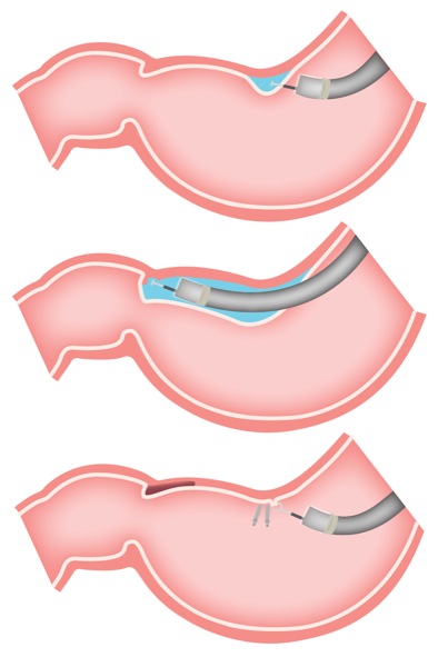
Image Source:HERE.Per-oral pyloromyotomy is a minimally invasive, lower-risk method of disrupting the pylorus that has been shown to improve gastroparesis symptoms.
The POP procedure was performed in the operating room under general anesthesia. Patients stayed in the hospital overnight after the POP procedure and an upper gastrointestinal (GI) series was performed to assess emptying through the pylorus and to check for unrecognized perforations. At discharge, patients were instructed to follow a liquid diet for two weeks and to take anti-acid therapies (sucralfate and a proton pump inhibitor) for four weeks. A repeat gastric-emptying study was performed at three months post-procedure.
The POP procedure produced statistically significant objective and subjective improvements in gastroparesis symptoms at 30 days and three months after treatment. Prior to the procedure, the average percentage of retained food at four hours was 37 percent and the average GCSI score was 4.6. After POP, the retained food percentage was reduced to 20 percent and the GCSI score to 3.3.
One patient died within 30 days of the POP procedure, but his death was unrelated to the surgery. “The procedure is very safe,” says Dr. Rodriguez, “and there were no procedure-related adverse events, including gastric or duodenal ulcer, intraluminal hemorrhage or gastric dumping syndrome.” There were also no repeat surgeries or hospitalizations related to the POP procedure.
A first-line treatment option
“This procedure has dramatically changed our practice at the Cleveland Clinic, and has become our first-line treatment option for medically refractive gastroparesis in well-selected patients,” Dr. Rodriguez says.
According to Matthew D. Kroh, MD, head of the research group and Chief of the Digestive Disease Institute at Cleveland Clinic Abu Dhabi, “POP is an attractive option for patients who in the past would have been offered surgical therapy because it is less invasive. This endoscopic procedure results that are similar to the best alternatives, without the morbidity associated with surgical access, and has a short recovery time.” Dr. Rodriguez adds that “because of its safety profile, we are able to apply POP more broadly than we could apply surgical interventions.”
Dr. Rodriguez reports that Cleveland Clinic has taken the lead in the U.S. with the POP procedure, but because GI motility is so complex and gastroparesis fairly common, he expects to see the technique become more widespread as more experience aggregates on its long-term safety and effectiveness."
"Early human experience with Per-Oral Endoscopic Pyloromyotomy (POP)
Eran Shlomovitz, MD, Radu Pescarus, MD, Ahmed Sharata, MD, Kevin M Reavis, MD, Christy M Dunst, MD, Lee L Swanstrom, MD. Providence Portland Medical Center, The Oregon Clinic..
Introduction:
Gastroparesis, a condition characterized by delayed gastric emptying, and a constellation of symptoms including nausea, vomiting, early satiety and bloating, is a debilitating condition. A variety of surgical options are available including pyloroplasty and pyloromyotomy. Although these have been shown to be effective they are associated with surgical trauma. We hypothesize that an endoscopic submucosal myotomy technique can be applied to endoscopically divide the pyloric sphincter, provide the benefits of a natural orifice procedure and improve gastric emptying in gastroparetic patients.
Methods and procedures:
Endoscopic per-oral pyloromyotomy (POP) was performed in four female patients ages 65, 59, 33 and 32 years old. All patient underwent a complete pre-operative work-up including upper endoscopy, gastric emptying study as well as a pH study and esophageal manometry if a concomitant fundoplication was performed. Three procedures were performed under laparoscopic guidance as patients required other concurrent laparoscopic procedures (see table). In one patient the procedure was fully endoscopic. The myotomy was performed by a technique similar to the one utilized in the POEM procedure. After the creation of a mucosotomy, a submucosal tunnel is established up to the duodenal bulb followed by a myotomy of the circular fibers of the pylorus. The mucosotomy is subsequently closed with clips.
Results:
Endoscopic per-oral pyloromyotomy was technically successful in all four cases and patients were discharged home on post operative day 2 or 3. There were no immediate procedural complications. One patient presented to the hospital 2 weeks post procedure with an upper GI bleed necessitating transfusions. On endoscopy a 1cm ulcer was found in the pyloric channel and an exposed vessel was clipped. The patient was subsequently discharged home on high dose proton pump inhibitors. Three month follow-up nuclear medicine gastric emptying studies (GES) are available for 3 of the 4 patients. Normalization of gastric emptying studies was demonstrated in 2 patients. Patient 3 showed improved gastric emptying half life, but unchanged residual activity at 4hrs.
Concomitant procedure
Operative time
Blood loss
Pre-op GES
Post-op GES
Patient 1
65 F
Cholecystectomy
102 min
Minimal
Half life: 150min
Residual at 4hrs: 29%
Half life: 36min
Residual at 4hrs: 0%
Patient 2
59 F
Redo- PEH repair and Nissen
295 min
100 cc
Half life: 90min
Residual at 4hrs: 14%
Half life: 18min
Residual at 4hrs: 0%
Patient 3
33 F
Nissen
231 min
Minimal
Half life: 160-170min
Residual at 4hrs: 15%
Half life: 70-90min
Residual at 4hrs: 14%
Conclusion:
Endoscopic pyloromyotomy is a technically feasible and potentially much less morbid endoscopic surgical procedure. Early follow-up suggests objective improvement in gastric emptying. Further long-term follow-up and additional clinical experience is required to establish the role of this technique in the management of gastroparesis."
Now, I want to discuss ERCP. I have had this procedure done to me, personally, and it helped me a great deal. I had my ERCP done in 2012, when I was first diagnosed with Gastroparesis. The doctors had to place a stent in my bile duct of my liver because it was not draining bile properly. You can read more about my personal experiences in my earlier blog articles:
I did develop pancreatitis after the procedure, which can be a side effect. That was a very painful experience. However, I want to research ERCP for those of you whose doctors may have suggested it, for those of you curious about it, and for those of you who might not know about the procedure itself.
There is a wonderful video with an explanation of the procedure made bySages,
This talk was presented at the 2018 SAGES Meeting/16th World Congress
of Endoscopic Surgery by Heidi J Miller during the When Bad Things
Happen to Good People – Endoscopy: Being FLEXible on April 14 2018
Endoscopic retrograde cholangiopancreatography (ERCP) is a procedure that combines upper gastrointestinal (GI) endoscopy and x-rays to treat problems of the bile and pancreatic ducts.
What are the bile and pancreatic ducts?
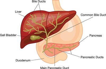
Your bile ducts are tubes that carry bile from your liver to your gallbladder and duodenum. Your pancreatic ducts are tubes that carry pancreatic juice from your pancreas to your duodenum. Small pancreatic ducts empty into the main pancreatic duct. Your common bile duct and main pancreatic duct join before emptying into your duodenum.
Why do doctors use ERCP?
Doctors use ERCP to treat problems of the bile and pancreatic ducts.
Doctors also use ERCP to diagnose problems of the bile and pancreatic
ducts if they expect to treat problems during the procedure. For
diagnosis alone, doctors may use noninvasive tests—tests that do not
physically enter the body—instead of ERCP. Noninvasive tests such as
magnetic resonance cholangiopancreatography (MRCP)—a type of magnetic resonance imaging (MRI) —are safer and can also diagnose many problems of the bile and pancreatic ducts.
Doctors perform ERCP when your bile or pancreatic ducts have become narrowed or blocked because of
gallstones that form in your gallbladder and become stuck in your common bile duct
To prepare for ERCP, talk with your doctor, arrange for a ride home, and follow your doctor’s instructions.
Talk with your doctor
You should talk with your doctor about any allergies and medical
conditions you have and all prescribed and over-the-counter medicines,
vitamins, and supplements you take, including
nonsteroidal anti-inflammatory drugs (NSAIDs) such as ibuprofen and naproxen
Your doctor may ask you to temporarily stop taking medicines that
affect blood clotting or interact with sedatives. You typically receive
sedatives during ERCP to help you relax and stay comfortable.
Tell your doctor if you are, or may be, pregnant. If you are pregnant
and need ERCP to treat a problem, the doctor performing the procedure
may make changes to protect the fetus from x-rays. Research has found
that ERCP is generally safe during pregnancy.1
Arrange for a ride home
For safety reasons, you can’t drive for 24 hours after ERCP, as the sedatives or anesthesia used during the procedure needs time to wear off. You will need to make plans for getting a ride home after ERCP.
Don’t eat, drink, smoke, or chew gum
To see your upper GI tract clearly, you doctor will most likely ask
you not to eat, drink, smoke, or chew gum during the 8 hours before
ERCP.
How do doctors perform ERCP?
Doctors who have specialized training in ERCP perform this procedure at a hospital or an outpatient center. An intravenous
(IV) needle will be placed in your arm to provide a sedative. Sedatives
help you stay relaxed and comfortable during the procedure. A health
care professional will give you a liquid anesthetic to gargle or will
spray anesthetic on the back of your throat. The anesthetic numbs your
throat and helps prevent gagging during the procedure. The health care
staff will monitor your vital signs and keep you as comfortable as
possible. In some cases, you may receive general anesthesia.
You’ll be asked to lie on an examination table. The doctor will carefully feed the endoscope down your esophagus, through your stomach,
and into your duodenum. A small camera mounted on the endoscope will
send a video image to a monitor. The endoscope pumps air into your
stomach and duodenum, making them easier to see.
During ERCP, the doctor
locates the opening where the bile and pancreatic ducts empty into the duodenum
slides a thin, flexible tube called a catheter through the endoscope and into the ducts
injects a special dye, also called contrast medium, into the ducts through the catheter to make the ducts more visible on x-rays
uses a type of x-ray imaging, called fluoroscopy, to examine the ducts and look for narrowed areas or blockages
The doctor may pass tiny tools through the endoscope to
insert stents—tiny tubes that a doctor leaves in narrowed ducts
to hold them open. A doctor may also insert temporary stents to stop
bile leaks that can occur after gallbladder surgery.
The procedure most often takes between 1 and 2 hours.
What should I expect after ERCP?
After ERCP, you can expect the following:
You will most often stay at the hospital or outpatient center
for 1 to 2 hours after the procedure so the sedation or anesthesia can
wear off. In some cases, you may need to stay overnight in the hospital
after ERCP.
You may have bloating or nausea for a short time after the procedure.
You may have a sore throat for 1 to 2 days.
You can go back to a normal diet once your swallowing has returned to normal.
You should rest at home for the remainder of the day.
Following the procedure, you—or a friend or family member who is with
you if you’re still groggy—will receive instructions on how to care for
yourself after the procedure. You should follow all instructions.
You will receive instructions on how to care for yourself after ERCP. You should follow all instructions.
Some results from ERCP are available right away after the procedure.
After the sedative has worn off, the doctor will share results with you
or, if you choose, with your friend or family member.
If the doctor performed a biopsy, a pathologist will examine the biopsy tissue. Biopsy results take a few days or longer to come back.
What are the risks of ERCP?
The risks of ERCP include complications such as the following:
pancreatitis
infection of the bile ducts or gallbladder
excessive bleeding, called hemorrhage
an abnormal reaction to the sedative, including respiratory or cardiac problems
perforation in the bile or pancreatic ducts, or in the duodenum near the opening where the bile and pancreatic ducts empty into it
tissue damage from x-ray exposure
death, although this complication is rare
Research has found that these complications occur in about 5 to 10 percent of ERCP procedures.2 People with complications often need treatment at a hospital.
As always, please discuss these procedures with your doctors. This is just research I have compiled but it does not take the place of a doctor's expertise or advice.
Endoscopic — Refers to a tool called an
endoscope, a long, thin (about the width of your little finger),
flexible tube with a camera on the end.
Retrograde — Refers to the direction (backward)
in which the endoscope injects a liquid for X-rays of parts of the GI
tract called the bile duct system and pancreas.
Cholangio — Refers to the bile duct system.
Pancreatography — Refers to the pancreas.
The process of taking these X-rays is known as cholangiopancreatography.
ERCP can help find the cause of jaundice (when your skin and/or the whites of your eyes turn yellow) or pancreatitis, which is inflammation (swelling and redness) of the pancreas that is often caused by gallstones or alcohol abuse. ERCP can also treat some of those issues.
Using tools passed through the endoscopic tube, your doctor can
inject dye to light up organs under X-rays. This provides a clear view
of your pancreas, pancreatic duct, bile duct system, gallbladder and
duodenum (the first portion of the small intestine).
ERCP is an endoscopic procedure used to inject dye into the bile and pancreas ducts. X-ray pictures are then taken.
ERCP can see if there is a blockage or narrowing in your biliary or pancreatic ducts caused by stones, tumors or scarring.
ERCP is frequently performed to find the cause of abnormal
liver-chemistry tests and to follow up on an abnormal ultrasound, CT
scan or MRI exam.
During an ERCP, if any blocks are found, tools can be passed through to relieve the block. Stones can be removed from the common bile duct or pancreatic duct and blocks can be dilated, biopsied and/or stented.
ERCP can relieve jaundice (when your skin and/or the whites of your eyes turn yellow) caused by blocked bile ducts.
ERCP can help find the cause of pancreatitis, inflammation (swelling and redness) of the pancreas, and prevent future attacks.
There are may different tests to confirm the diagnosis of Gastroparesis. I wanted to write an article to list the different methods doctors can use to determine whether or not you have Gastroparesis. Sometimes, multiple tests are needed. Speak with your doctor before you are tested, just in case you are on any medications which can skew the test results (i.e. opiates like hydrocodone can slow down motility and prevent you from receiving an accurate diagnosis.
The first test I want to discuss is the breath test, which can be used to determine if someone has Gastroparesis or not. I have never been tested using the breath test method, so I researched the topic to understand it as well as to write about it.
Testing for Gastroparesis: The Breath Test
According to the US PHARMACIST,
"Gastric Emptying Breath Test
Kendra L. Sutton, PharmD Candidate
Florida A&M University
College of Pharmacy
Tallahassee, Florida
Cy D. Sims, PharmD Candidate
Florida A&M University
College of Pharmacy
Tallahassee, Florida
Marlon S. Honeywell, PharmD
Professor of Pharmacy Practice
Florida A&M University
College of Pharmacy
Tallahassee, Florida
Ellen Campbell, PhD
Associate Professor of Economic, Social and Administrative Pharmacy
Florida A&M University College of Pharmacy and Pharmaceutical Sciences
Tallahassee, Florida
Tracy A. Thomas, PT, PhD
Associate Professor of Physical Therapy
Florida A&M University
Tallahassee, Florida
Eboni Allen, MSN, ARNP-C
Bond Community Health Center
Tallahassee, Florida
Anneka J. Gaffney, MSN, ARNP-BC
Bond Community Health Center
Tallahassee, Florida
Temple Robinson, MD
Chief Medical Officer
Bond Community Health Center
Tallahassee, Florida
U.S. Pharm. 2015;40(12):HS36-HS40.
Delayed gastric emptying, also known as gastroparesis, is a disorder that either slows or stops movement of food through the gastrointestinal (GI) tract. It commonly causes nausea, vomiting, heartburn, erratic blood glucose levels, and postprandial fullness.1 Normally, the smooth muscles of the GI tract move food down to the stomach and through the intestines, where it is excreted through the rectum. This process is controlled by the vagus nerve, a component of the parasympathetic nervous system. The most common causes of gastroparesis are diabetes mellitus and surgery; it may also be of unknown cause. Diabetes accounts for almost one-third of cases of gastroparesis, with about 5% to 12% of all diabetes patients experiencing this disorder.2,3 Diagnosis is most often determined by performing endoscopy or using scintigraphy—a specialized radiologic imaging technique—to measure the rate at which radioactive food passes through the GI tract. The Gastric Emptying Breath Test (GEBT) makes it possible to diagnose gastroparesis without the use of radioactive materials.
Gastric Emptying Breath Test
Developed by Advanced Breath Diagnostics, the GEBT is a non-radioactive test that utilizes carbon-13 (13C). Labeling with 13C stable isotope is essentially safe, as 1.1% of our bodies, and of the food we eat, consists of 13C; the remaining 98.9% consists of 12C.4 The GEBT test was developed for adult patients who are symptomatic for gastroparesis. The test measures the rate of gastric emptying of solids and aids in the diagnosis of gastroparesis. The test system utilizes a gas isotope ratio mass spectrometer for the measurement of the ratio of 13CO2 to 12CO2 in breath samples. Administration of the GEBT does not require a special facility, but it should be administered under the guidance of a healthcare professional.4 The GEBT is shown in FIGURE 1.
Source: in article
Test Administration
The GEBT is measured over a 4-hour time cycle. Detailed administration instructions can be found in Table 1. Multiple breath samples are obtained from the patient; sampling should be performed subsequent to an 8-hour fast. After providing multiple premeal breath samples (Table 2), the patient consumes the standard GEBT meal consisting of pasteurized scrambled egg mix containing a dose of 43 mg of 13C-Spirulina, 6 saltine crackers, and 6 ounces of potable water.5 The 13C-Spirulina is eventually passed through the GI tract to the intestines; it is in the intestines that the Spirulina is absorbed and metabolized to the 13CO2 expired in the breath. Single postmeal breath samples are subsequently collected (Table 2) at 45, 90, 120, 150, 180, and 240 minutes from the end of test-meal consumption. The post-meal breath samples are transmitted to a laboratory for assessment of 13CO2/12CO2 in each sample. By measuring the change in this ratio over time as compared to the pre-meal value, the rate of 13CO2 excretion can be calculated and the patient’s gastric-emptying rate determined.4,5 See FIGURE 2 for a diagram outlining the procedure.
Source: in article
Efficacy
Scintigraphy (The definition of Scintigraphy, according to the Dictionary,
"noun, MEDICINE. A technique in which a scintillation counter or similar detector is used with a radioactive tracer to obtain an image of a bodily organ or a record of its functioning."), a competitor to the GEBT, utilizes a radioisotope that is unsafe for patients who are having multiple tests performed, pregnant women, women who breast-feed, and children. Scintigraphy also requires special equipment and a specific testing site, whereas the GEBT can be implemented outside the clinical setting and breath samples are subsequently sent to a laboratory for analysis.4-6
In a clinical trial comparing the GEBT and scintigraphy, the GEBT was shown to be comparable to scintigraphy. Researchers compared the results from both the GEBT and scintigraphy and found that GEBT results agreed with scintigraphy results 73% to 97% of the time when measured from various time points during the test.4,5 The GEBT was also found to be safer than scintigraphy.
Adverse Effects/Contraindications
In the comparative study mentioned above, 13 participants complained of adverse effects.5,7 Adverse events experienced were often considered mild; these events included nausea, heartburn, diarrhea, dry heaves, abdominal pain, acid reflux, dizziness, and head cold. It was also determined later that many of these events were not related to the device. Contraindications to use of the GEBT include egg, milk, or wheat allergy, as the food product ingested contains these ingredients.5,8 Additionally, as the digestion of the product involves other organs such as the pancreas and liver, the GEBT is not recommended in patients with malabsorption, or who have diseases such as pancreatitis or hepatitis.
Conclusion
The GEBT is unique in that it provides a nonradioactive, noninvasive, orally administered product that performs as well as scintigraphy. The ease of use outside of the clinical setting also makes this device an attractive alternative to other available tests. For more information, contact Advanced Breath Diagnostics at (615) 376-5464.
REFERENCES
1. National Institutes of Health. National Institute of Diabetes and Digestive and Kidney Diseases. Gastroparesis. June 2012. www.niddk.nih.gov/health-information/health-topics/digestive-diseases/gastroparesis/Pages/facts.aspx. Accessed July 10, 2015.
2. Soykan I, Sivri B, Sarosiek I, et al. Demography, clinical characteristics, psychological and abuse profiles, treatment, and long-term follow-up of patients with gastroparesis. Dig Dis Sci. 1998;43:2398-2404.
3. Bytzer P, Talley NJ, Leemon M, et al. Prevalence of gastrointestinal symptoms associated with diabetes mellitus: a population-based survey of 15,000 adults. Arch Intern Med. 2001;161:1989-1996.
4. Szarka L, Camilleri M, Vella A, et al. A stable isotope breath test with a standard meal for abnormal gastric emptying of solids in the clinic and in research. Clin Gastroenterol Hepatol. 2008;6:635-643.
5. FDA. Gastric emptying breath test. www.accessdata.fda.gov/cdrh_docs/pdf11/P110015b.pdf. Accessed July 21, 2015.
6. Parkman HP, Hasler WL, Fisher RS. American Gastroenterological Association medical position statement: diagnosis and treatment of gastroparesis. Gastroenterology. 2004;127:1589-1591.
7. Abell TL, Bernstein RK, Cutts T, et al. The American Motility Society Task Force on Gastroparesis. Treatment of gastroparesis: a multidisciplinary clinical review. Neurogastroenterol Motil. 2006;4:263-283.
8. Park M, Camilleri M. Gastroparesis: clinical update. Am J Gastroenterol. 2006;101:1129-1139.
9. Gastric Emptying Breath Test [package insert]. Brentwod, TN: Advanced Breath Diagnostics, LLC; 2015.
To comment on this article, contact rdavidson@uspharmacist.com."
"The FDA recently announced the approval of the Gastric Emptying Breath Test (GEBT) as a diagnostic tool for gastroparesis – this condition is, though not as well known, a common complication of diabetes. Gastroparesis slows or stops the movement of food down the digestive tract from the stomach to the small intestine – this is caused by problems with stomach muscle contractions, which usually help move the food forward. In other words, digestion doesn't go nearly as well as it should. The GEBT works by measuring carbon dioxide in a patient’s breath over a four-hour period after eating a 'test meal.' The carbon dioxide measurement is used to calculate the rate at which food is emptied from the stomach.
GEBT should offer a major improvement over the current gold standard for gastroparesis diagnosis, gastric emptying scintigraphy. Gastric emptying scintigraphy requires eating radioactively labeled foods and measuring their digestion with an external scanner. As the GEBT does not require imaging equipment or training for the handling of radioactive materials, the hope is that it will be easier for more health care providers to use in their practices. A clinical study of 115 people conducted to support GEBT’s approval demonstrated that results from the GEBT agreed with the results of scintigraphy 73%-97% of the time.
Gastroparesis is a fairly common complication of diabetes, impacting an estimated 40% of type 1 and 30% of type 2 patients. In addition to causing symptoms like nausea and heartburn (see a full list of symptoms here), gastroparesis can make managing diabetes much more difficult, as it can lead to increased blood glucose variability and hypoglycemia. Gastroparesis remains an area of significant need, as no new therapies have been approved in the US in over 30 years and dietary interventions have not been clearly demonstrated to work. The approval of the GEBT is a positive step towards increased detection of this complication; for more information on diabetes and gastroparesis, please see the ADA’s page on it here. –AJW/ER"
I have already written about this before (in 2012), but was speaking about my own experiences with The Smart Pill Test. At the time I wrote this article, there was not a lot of information online about Gastroparesis, much less the different tests used to diagnose someone who has Gastroparesis.
Therefore, I wanted to put all of the testing I am aware of, to determine Gastroparesis, in the same article. The link to my article about The Smart Pill Test can be found here: http://www.emilysstomach.com/2012/08/smart-pill.html
Furthermore, GASTRO CURE says this about The Smart Pill,
"GI MONITORING SYSTEM
SmartPill is an ingestible, wireless capsule that measures pressure, pH and temperature as it transits the GI tract. This information is used to provide regional transit times including gastric emptying time, small bowel transit time, colonic transit time, combined small/large bowel transit time, whole gut transit time, pressure contraction patterns from the antrum and duodenum and motility indices.
Administered in the physician’s office, the SmartPill test is completely ambulatory and allows the patient to go about their normal routine during the course of the test. As the SmartPill Capsule passes through the GI tract, it transmits data to a SmartPill Data Receiver worn by the patient. Once the single-use capsule has passed from the body, the patient returns the data receiver to the Physician who then can download the collected data to a PC. The Physician then uses SmartPill’s MotiliGI software to display and analyze the data, providing the physician with test results in both graphical and report formats.
The SmartPill Wireless Motility Capsule
The SmartPill Capsule transits the intestines by peristalsis, or the normal rhythmic contraction of the intestinal muscles, and is capable of transmitting data continuously for at least five days. The single-use capsule is excreted naturally from the body, usually within a day or two.
SmartPill Data Receiver & Docking Station
The battery-operated SmartPill Data Receiver collects and stores the test data. The Receiver is capable of transmitting data for real-time monitoring of a test. It also stores a complete record of the data captured by the SmartPill Capsule for download to a PC after the test is complete.
The SmartPill Data Receiver must be worn by the patient for the duration of the test on a belt clip or a lanyard around their neck. The data receiver may be removed for sleeping or bathing but should be kept in close proximity at all times.
After completion of the test, the SmartPill Data Receiver is connected to the system computer using the SmartPill Docking Station. The docking station features an industry standard USB type 2.0 interface that is used to transfer captured data to the PC for analysis and data reconstruction using SmartPill’s MotiliGI software. The data analysis can be performed at the convenience of the physician in a single session or during multiple sessions without the need for the patient to be present.
SmartPill MotiliGI Software
SmartPill MotiliGI software comes pre-installed on the system PC and provides an graphical user interface. MotiliGI receives and processes data from the SmartPill Data Receiver, stores the data in a file on the system PC, provides a test summary and report, graphs for display purposes, and shows the data values captured during the SmartPill test. MotiliGI provides powerful data analysis tools, and affords the user a variety of test reporting and exporting options.
System Computer
The SmartPill GI Monitoring System is supplied with a personal computer which hosts MotiliGI software and supports communication between MotiliGI and the data receiver and docking station.
SmartPill Activation Fixture
The activation fixture contains very powerful magnets and is used to activate and deactivate the SmartPill Capsule."
I wanted to update this a bit since there is A LOT more information about The Smart Pill Test than when I was first diagnosed. For instance, I did not know that The Smart Pill Test could not work with certain conditions, like Crohn's Disease. So, I have actually learned a lot of new things about The Smart Pill test that I was unaware of before.
"Gastroparesis: ‘Smart’ Pill Uncovers This Mysterious Stomach Condition: Data-Gathering Pill Moves Through Your GI Tract
If you have unexplained stomach problems, such as nausea, bloating, constipation, chronic abdominal pain or vomiting, you might swallow a pill — but not the medicated kind.
Instead, consider a 'smart' pill, which gathers data as it moves through your GI tract. This painless tool helps your doctor pinpoint the cause of your problem so you can get the best treatment, says gastroenterologist Michael Cline, DO.
How can the pill help?
Data gathered by such a pill could help your doctor diagnose a condition called gastroparesis, which causes food to move abnormally slowly from your stomach to the small intestine, while ruling out another GI condition, such as chronic constipation.
Gastroparesis, which means partial paralysis of the stomach, is a common condition for those with diabetes. The condition reduces your stomach’s ability to empty its contents, but it does not involve a blockage.
'Basically, gastroparesis is a stomach that doesn’t empty well,' says Dr. Cline.
And it’s a difficult disease to treat, he says. Treatment typically begins with adjustments to diet and medication. If those approaches don’t work, surgical treatments are the next steps.
How does the pill work?
The SmartPill® provides a simple, painless way to collect data about your GI tract without using radiation, Dr. Cline says. It’s an FDA-approved, disposable capsule that you swallow.
'It’s called ‘smart’ because it measures the acid level, the pressure level, the temperature and time of your GI tract,' he explains. 'It’s similar to the pill cam, which takes pictures inside your body. This is its sister, essentially.'
Instead of taking photos of the inside of your body, the SmartPill measures motility and movement of your gut. It wirelessly transmits data about your GI tract to a recorder that you wear on a belt clip or lanyard around your neck as you go about your daily activities.
'The recorder captures the data and we download it into a computer and print out what’s happening, from start to finish,' Dr. Cline says. 'We can get all of this data and measurements of motility and movement without any wires attached and without X-rays.'
Depending on how slowly your intestines move, the SmartPill typically works its way through your system in three to five days. If you have normal motility, it is in your stomach for less than four hours, in your small intestine for less than six hours and in your large intestine or colon for less than 59 hours.
The SmartPill can also help a physician rule out other gastric issues that can cause problems in your small intestine or colon.
'You want to make sure that you’re dealing with just gastroparesis and not another issue,' says Dr. Cline.
The one real benefit of the SmartPill over everything else available is that it measures all three sections of your intestine with one test. Dr. Cline says that not only are you looking for gastroparesis, but you’re also looking at your small intestine movement and colon movement.
'There’s no other test out there that can measure all three with one test,' he says. 'I tell patients you get a lot more bang for your buck with the SmartPill.'
Is a ‘smart’ pill right for you?
The SmartPill does not work for people with certain conditions, Dr. Cline says. These conditions include:
A history of strictures in your intestines, such as scarring (which can narrow the bowel passage)
Inflammatory bowel disease, like Crohn’s disease
A heart pacemaker (or pacemakers in other organs)
'The main risk of the SmartPill is it could get stuck,' he explains. 'If you have normal intestines, the SmartPill will not stick. If the pill gets stuck, you have a problem, obviously. You don’t want to give a patient who is at a high risk of having narrowing bowels the SmartPill.'
The FDA advises against giving the SmartPill to patients who have pacemakers.
Dr. Cline says he also advises patients who need an MRI to delay taking the SmartPill.
'The final contraindication, which is pretty rare, is if the patient has an MRI scheduled,” he says. 'You obviously don’t want to do it until after the MRI is over. You can’t go through an MRI with that pill inside of you.'"
For more information about The Smart Pill, please visit:
A gastric emptying scan (GES) is a nuclear medicine exam that uses a radioactive material that you will eat in a meal. You will eat this meal in the Radiology department before your scan. The radioactive material allows doctors to see how your stomach empties.
This scan is used to help diagnose conditions called motility disorders. These are conditions that change the way the stomach contracts and moves food into your intestines. A GES is a form of radiology, because radiation is used to take pictures of your body.
How does the scan work?
A gamma camera takes pictures as the radioactive food moves through your stomach. The camera detects the gamma rays emitted from the food. A computer then produces pictures and measurements of your stomach.
How should I prepare for the scan?
You will need to fast for 6 hours before the scan.
If you smoke, do not smoke the morning of the test and during the hours of the scan.
If you are a woman and you are still menstruating, it is best to have your scan done in the first 10 days of your menstrual cycle.
Do not take these drugs for at least 2 days before your test, unless your doctor tells you otherwise:
Drugs called prokinetic agents that speed up the motility of your GI tract. Some of these are metoclopramide (Reglan), erythromycin, tegaserod (Zelnorm), and domperidone (Motilium).
Drugs called anticholinergic antispasmodic agents that slow down the motility of your GI tract. Some of these are Bentyl, Donnatal, Levsin, Robinul, and Hyosyne.
Pain medicines called opiate analgesics. Some of these are codeine, demerol, Percocet (Oxycodone), Tylenol #3, Tylox, Oxycontin, Percodan, Fentanyl patch, morphine, Methadone, Vicodin (Hydrocodone), and Ultram (Tramadol).
Marijuana.
Do not take any laxatives the day before or any time during the test.
Note: It is OK to take relaxants(called benzodiazepines)such as Ativan, Valium, Librium, Xanax, and others. It is also OK to take anti-nausea medicines (called phenothiazines) such as Thorazine, Compazine, Phenergan, and others. These drugs do not affect gastric emptying.
You may take all other medicines the day of your test with small sips of water. Take these at least 2 hours before your test begins.
If you have severe nausea on the day of your test, we may give you medicine to reduce your nausea.
If You Have Diabetes
On the day of the test:
Your fasting blood glucose should be less than 275 mg/dL. A technologist will check your blood glucose before your test starts.
Please bring your insulin or other diabetes medicine with you. You may need to take it with your gastric emptying meal.
How is the scan done?
You will be given 2 small sandwiches, one filled with jam and one filled with 4 cooked egg whites that have been injected with a small dose of radioactive material. You will be asked to eat the sandwiches and drink water within 10 minutes. The egg will taste just like a regular egg. If you are allergic to eggs or wheat, please tell the person you schedule your appointment with. A different meal will be used.
After eating, you will be asked to lie flat on your back while the gamma camera take a picture of your stomach. You must lie still when the camera is taking pictures. If you move, the pictures will be blurry and may have to be taken again.
What will I feel during the scan?
Lying still on the exam table may be hard for some patients. The technologist will help make you comfortable.
How long will the scan take?
From start to finish, your gastric emptying scan will take about 4 hours.
There are 4 parts to the test:
First half hour: Eat meal, then take pictures with gamma camera (pictures take 5 minutes)
1 hour after meal: Take pictures (5 minutes)
2 hours after meal: Take pictures (5 minutes)
4 hours after meal: Take pictures (5 minutes)
After eating the meal:
You may leave the Nuclear Medicine department between the times you have the pictures taken.
Do not eat or exercise until after the last set of pictures has been taken.
Who interprets the results and how do I get them?
When the test is over, the nuclear medicine doctor will review your images, write up a report, and talk with your doctor about the results. Your doctor will talk with you about the results and your treatment options."
Source: My own photo
According to HEALTH LINE (which has great information about the test and alternatives) also has information about Gastric Emptying Scans for children,
"Gastroparesis symptoms in children are similar to those seen in adults. Ask your doctor to administer this test to your child if they’re experiencing any of symptoms mentioned earlier.
The test for older children is identical to the test given to adults. If your child is a baby or infant, your doctor gives your child the radioactive food in milk or formula in an exam known as a milk study or liquid study. In this case, you may be instructed to bring your own formula or milk from home to make sure your child doesn't have an allergic reaction.
The radioactive substance is just as safe for your child as it is for an adult. The test usually takes about three hours for children. If your child is given the liquid study instead, the camera takes continuous images for about an hour. It’s important that your child remains still throughout the test. Make sure that you find a way to keep them occupied or calm before and during the test so that the results can be delivered smoothly.
The following items may help keep your child relaxed:
music
toys
movies
books
comfort objects, such blankets or pillows"
The Test will accurately verify the presence of a common Digestive Disorder
The Physician will require a complete Medical History before the Test
There will be a Transceiver that is placed over the Patient’s stomach during the test.
The pre test instructions will help achieve the most accurate results.
There is no discomfort during a Heidelberg pH test. Patients are relaxed and comfortable.
Remember to grab a good old fashioned distraction to pass the time.
Due to wireless interference, cell phones and personal electronic devices may need to be powered off.
The results are immediate so the Physician may have the post test consultation the same day.
Heidelberg pH Diagnostic Test will accurately verify the presence of low stomach acid production, high stomach acid production, no acid in the stomach, Dumping Syndrome, Acute or sub Acute Gastritis, Heavy Mucus in the stomach, and Pyloric insufficiency.
The Test is accomplished by measuring the time it takes for the acid producing cells (Parietal Cells) in the stomach to produce the required Hydrochloric acid. The strength of the acid (pH) is also measured during the test.
Patients are required to complete a consent form for the Physician’s Office Staff. If you have reservations about having a pH test, please discuss them with the Physician. Mental anguish and stress will only alter the test results.
The Technician will calibrate the pH Capsule to ensure it accurately records the pH values. The Technician will then place a transceiver over the Patient’s stomach. The transceiver receives and transmits the information from the pH Capsule and sends it wirelessly to the computer where the information is displayed. The nurse will rinse the pH Capsule and give it the Patient to swallow with a sip of water.
There is no discomfort during a Heidelberg pH Test. Patients are asked to relax and get comfortable while the test is being done. Remember to grab a good old fashioned distraction to pass the time.
Results are ready for the Physician to review as soon as the Technician ends the Test. Many Physicians opt to have a post test consultation as soon as the test is complete. In many cases the doctor will establish a treatment protocol on the same day. In many instances, after testing and treatment, patients stated that they feel better and healthier in just two or three days.
Unlike other procedures, the Heidelberg pH diagnostic test is an in-office procedure that does not require sedation or the use of a stomach tube.
There is no trauma or discomfort associated with our test, and the results of the Heidelberg test are available to the doctor as soon as the test is complete."
I have had this test done as well. After I was first diagnosed with Gastroparesis, my doctor sent me to the Mayo Clinic to meet with a motility specialist there. I had to go off all of my other medications for a month before the doctor at Mayo would even see me, as I have mentioned before.
My personal experience was in February of 2013, and you can read the article here:
But, I want to discuss the test because the test now is a bit different than when I took it in 2013. Even though it is used as testing for GERD, my personal test found fifty-five events in twenty-four hours, with forty-six of them being vomiting. So, it might measure for GERD but my test was used to prove my vomiting with Gastroparesis, and how much I vomit.
A small capsule, about the size of a gel cap, is temporarily attached to the wall of the esophagus during an upper endoscopy. The capsule measures pH levels in the esophagus and transmits readings by radio telecommunications to a receiver (about the size of a pager) worn on your belt or waistband. The receiver has several buttons on it that you will press to record symptoms of GERD such as heartburn (the nurse will tell you what symptoms to record). You will be asked to maintain a diary to record certain events such as when you start and stop eating and drinking, when you lie down, and when you get back up. This will be explained by the nurse.
How do I prepare for the Bravo esophageal pH test?
Let your physician know if you have a pacemaker or implantable heart defibrillator, a history of bleeding problems, dilated blood vessels, and any other previously known problems with your esophagus.
Seven days before the monitoring period, do not take proton pump inhibitors such as omeprazole (Prilosec®), lansoprazole (Prevacid®), rabeprazole (Aciphex®), pantoprazole (Protonix®), esomeprazole (Nexium®).
Two days (48 hours) before the monitoring period, do not take the H2 blockers ranitidine (Zantac®), cimetidine (Tagamet®), famotidine (Pepcid®), nizatidine (Axid®); or the promotility drug, metoclopramide (Reglan®).
Six hours before the monitoring period, do not take antacids (such as Alka-Seltzer®, Gaviscon®, Maalox®, Milk of Magnesia®, Mylanta®, Phillips®, Riopan®, Tums® or any other brands).
Four to 6 hours before your appointment do not eat or drink.
Please note: Occasionally, your doctor may want you to continue taking a certain medication during the monitoring period to determine if it is effective.
Once the test has begun, what do I need to know and do?
Activity: Follow your usual daily routine. Do not reduce or change your activities during the monitoring period. Doing so can make the monitoring results less useful. Note: Do not get the receiver wet; it is not waterproof!
Eating: Eat your regular meals at the usual times. If you do not eat during the monitoring period, your stomach will not produce acid as usual, and the test results will not be accurate. Eat at least 2 meals a day. Eat foods that tend to increase your symptoms (without making yourself miserable). Avoid snacking. Do not suck on hard candy or lozenges and do not chew gum during the monitoring period.
Lying down: Remain upright throughout the day. Do not lie down until you go to bed (unless napping or lying down during the day is part of your daily routine).
Medications: Continue to follow your doctor’s advice regarding medications to avoid during the monitoring period.
Recording symptoms: Press the appropriate button on the receiver when symptoms occur (as discussed with the nurse). Record the time you start and stop eating and drinking (anything other than plain water). Record the time you lie down (even if just resting) and when you get back up. The nurse will explain this.
Unusual symptoms or side effects: If you think you may be experiencing any unusual symptoms or side effects, call your doctor.
You will return the receiver and diary when the monitoring period is over. The information on the receiver and diary will be downloaded to a computer and the results will be analyzed. Preliminary study data will be reviewed with you if you desire.
After completion of the study:
Resume your normal diet and medications.
Your doctor will discuss the results of your test with you during your next scheduled appointment.
Normal activities – such as swallowing, eating and drinking – will cause the disposable pH capsule to detach and pass through the digestive tract in 7 to 10 days on average.
No MRI exams (magnetic resonance imaging) should be performed for 30 days following capsule insertion."
These are the five main tests that are preformed to properly diagnose Gastroparesis. Doctors may use a combination of these tests to determine if:
1. You have Gastroparesis
2. How mild to severe your Gastroparesis is
I did not really get into endoscopies and upper GI testing, like barium swallows, because those are not the best tests to determine whether or not someone has Gastroparesis. These tests can definitely be useful, and I am not debating that, but there are a lot of things that can missed on tests like these. My GI did an endoscopy on me, before he hospitalized me to do more testing (before I even knew what Gastroparesis was) and when I came to, he started getting on to me about eating after midnight and not telling them. I swore to him that I had not eaten or had anything to drink after midnight. I told him the last thing I ate, which was three days before the endoscopy, was a salad. He was appalled because I still had undigested salad greens sitting in my stomach. After that, he hospitalized me and did a gastric emptying study. At the end of the day, endoscopies and barium swallows can help confirm a diagnosis, but the other tests are a lot more accurate in diagnosing Gastroparesis, in my opinion, but you should talk with your doctor about the different test options if you suspect that you have Gastroparesis, or if a family member does.
As most of you probably already know, one of the experimental treatments for Gastroparesis is a Botox injection into the stomach through endoscopy.
According to the Mayo Clinic, Botulinum toxin A (BOTOX) is a powerful inhibitor of muscle contraction. Botox is a nerve toxin most commonly known for its use in treating skin wrinkles but is experimental for Gastroparesis. Botox can be injected through an endoscope to relax muscles in the GI tract, including the lower esophageal sphincter (end of the esophagus) in achalasia, and the pylorus (end of the stomach) in patients with Gastroparesis. Researchers have found that Botox injections relax the pyloric muscle in some people, thereby allowing the stomach to release more food into the small intestine. The benefits are temporary, however, and more studies are needed to determine the overall usefulness of this treatment.
I've read about it, did research on it, and asked people I knew who had it done questions about the procedure and if they would recommend it. Everyone I talked to had a great experience with it, so I decided that it couldn't hurt. Besides that, I was miserable and desperate.
So, I'm going to walk you through the botox injection procedure, step by step in what I went through today.
I have to admit I was nervous when I arrived at the hospital because I didn't know what to expect since I've never done this before. As soon as I walked in, I checked into the GI department. After I registered, I sat in the waiting room for a while.
The nurse called me back and put my fancy ID bracelet on. She gave me a gown and told me to put all of my stuff in a plastic bag. I had to strip down to my underwear for the hospital gown. After I was ready, she came back in and placed my plastic bag under my stretcher so that it could ride with me. She also logged all of my valuables.
She went ahead and applied the electrodes to monitor my heart rhythm, applied a blood pressure, and an oxygen sensor to take my vital signs. She tried to stick me for an IV, but missed the vein. So, they had to call in the IV team. Thankfully, the nurse on the IV team got me on the first try but had to use a really tiny needle for the IV because of my tiny veins. I was actually impressed because it usually takes them about six tries before they get it right.
The nurse gave me Zofran through the IV because I was extremely nauseated (since I couldn't eat or drink after midnight nor take my medications) while I was waiting.
I had a visit from the anesthesiologist and the doctor to make sure I didn't have any allergies, adverse reactions, etc.
Then, it was time to take me back for the endoscopy. If you've never had an endoscopy, the doctor uses an instrument to examine the interior of a hollow organ or cavity of the body. Unlike most other medical imaging devices, endoscopes are inserted directly into the organ and in this case, my stomach.
Below is an endoscope, image from Wikipedia:
Once I was in the room, the nurse had me lay on my side, facing away from the screen that the camera projects on to. She put oxygen in my nose and told me to open my mouth and bite down on a solid green circle, which would hold my mouth open for the camera to pass through.
The top picture is the bite block for an endoscope and the bottom picture is where the bite block goes. Source HERE.
The anesthesiologist then began to push the medications to put me to sleep for the endoscope. The first medication he pushed into my IV made my ears ring like crazy and made me a bit dizzy. The second one he told me would burn, and it did! It burned all of the way up my arm, so much so that I had tears in my eyes. It didn't hurt for very long because I woke up in recovery after that.
In recovery, the doctor visited my husband and myself. He said that if this doesn't help me, he would refer me to Augusta to a motility specialist there. After he left the room, the nurse came in, took my vital signs, and readied my discharge papers. She disconnected my IV, I got dressed, and she wheeled me out to my husband in our waiting car. That's all there was to it!
I was still groggy when I got home so I slept most of the day. I will tell you that I'm having stomach spasms because of the injection today. I was told it would take a few days to subside. So, I will keep you updated on my progress. In the meantime, check out some pictures my husband took of me in recovery. I look so thrilled!