Find us on Google+ Gastroparesis: stomach

Copyright

“You agree that you will not modify, copy, reproduce, sell, or distribute any content in any manner or medium without permission."
Showing posts with label stomach. Show all posts

Friday, September 22, 2017

The Brain in Your Gut

I know a lot of people with gastroparesis have memory issues, myself included. So, what causes this? How does our brain chemistry change when you have gastroparesis? Sleep deprivation, malnutrition, and medication can play a big part in altering our brain chemistry but I wanted to dig a bit deeper to see what else can change our body's brain chemistry, and why it affects us so harshly.


Credit: ISTOCKPHOTO/ERAXION


According to Cal Tech http://www.caltech.edu/news/microbes-help-produce-serotonin-gut-46495,

"Although serotonin is well known as a brain neurotransmitter, it is estimated that 90 percent of the body's serotonin is made in the digestive tract. In fact, altered levels of this peripheral serotonin have been linked to diseases such as irritable bowel syndrome, cardiovascular disease, and osteoporosis.

'More and more studies are showing that mice or other model organisms with changes in their gut microbes exhibit altered behaviors,' explains Elaine Hsiao, research assistant professor of biology and biological engineering and senior author of the study. 'We are interested in how microbes communicate with the nervous system. To start, we explored the idea that normal gut microbes could influence levels of neurotransmitters in their hosts.'

Peripheral serotonin is produced in the digestive tract by enterochromaffin (EC) cells and also by particular types of immune cells and neurons. Hsiao and her colleagues first wanted to know if gut microbes have any effect on serotonin production in the gut and, if so, in which types of cells. They began by measuring peripheral serotonin levels in mice with normal populations of gut bacteria and also in germ-free mice that lack these resident microbes.

The researchers found that the EC cells from germ-free mice produced approximately 60 percent less serotonin than did their peers with conventional bacterial colonies. When these germ-free mice were recolonized with normal gut microbes, the serotonin levels went back up—showing that the deficit in serotonin can be reversed.

'EC cells are rich sources of serotonin in the gut. What we saw in this experiment is that they appear to depend on microbes to make serotonin—or at least a large portion of it,' says Jessica Yano, first author on the paper and a research technician working with Hsiao.

The researchers next wanted to find out whether specific species of bacteria, out of the diverse pool of microbes that inhabit the gut, are interacting with EC cells to make serotonin.

After testing several different single species and groups of known gut microbes, Yano, Hsiao, and colleagues observed that one condition—the presence of a group of approximately 20 species of spore-forming bacteria—elevated serotonin levels in germ-free mice. The mice treated with this group also showed an increase in gastrointestinal motility compared to their germ-free counterparts, and changes in the activation of blood platelets, which are known to use serotonin to promote clotting.

Wanting to home in on mechanisms that could be involved in this interesting collaboration between microbe and host, the researchers began looking for molecules that might be key. They identified several particular metabolites—products of the microbes' metabolism—that were regulated by spore-forming bacteria and that elevated serotonin from EC cells in culture. Furthermore, increasing these metabolites in germ-free mice increased their serotonin levels.

Previous work in the field indicated that some bacteria can make serotonin all by themselves. However, this new study suggests that much of the body's serotonin relies on particular bacteria that interact with the host to produce serotonin, says Yano. 'Our work demonstrates that microbes normally present in the gut stimulate host intestinal cells to produce serotonin,' she explains.

'While the connections between the microbiome and the immune and metabolic systems are well appreciated, research into the role gut microbes play in shaping the nervous system is an exciting frontier in the biological sciences,' says Sarkis K. Mazmanian, Luis B. and Nelly Soux Professor of Microbiology and a coauthor on the study. 'This work elegantly extends previous seminal research from Caltech in this emerging field'.

Additional coauthor Rustem Ismagilov, the Ethel Wilson Bowles and Robert Bowles Professor of Chemistry and Chemical Engineering, adds, 'This work illustrates both the richness of chemical interactions between the hosts and their microbial communities, and Dr. Hsiao's scientific breadth and acumen in leading this work.'

Serotonin is important for many aspects of human health
, but Hsiao cautions that much more research is needed before any of these findings can be translated to the clinic.

'We identified a group of bacteria that, aside from increasing serotonin, likely has other effects yet to be explored,' she says. 'Also, there are conditions where an excess of peripheral serotonin appears to be detrimental.'

Although this study was limited to serotonin in the gut, Hsiao and her team are now investigating how this mechanism might also be important for the developing brain. 'Serotonin is an important neurotransmitter and hormone that is involved in a variety of biological processes. The finding that gut microbes modulate serotonin levels raises the interesting prospect of using them to drive changes in biology,' says Hsiao.

The work was published in an article titled 'Indigenous Bacteria from the Gut Microbiota Regulate Host Serotonin Biosynthesis.' In addition to Hsiao, Yano, Mazmanian, and Ismagilov, other Caltech coauthors include undergraduates Kristie Yu, Gauri Shastri, and Phoebe Ann; graduate student Gregory Donaldson; postdoctoral scholar Liang Ma. Additional coauthor Cathryn Nagler is from the University of Chicago."


Image Credit: http://i2.wp.com/sitn.hms.harvard.edu/wp-content/uploads/2016/08/Gut-Brain-Microbe-Figures_FINAL.png


This is an interesting study considering that Gastroparesis/DTP is slow to little to no motility, depending on how severe it is in each person affected with it. If 90 percent of serotonin is produced in the stomach, what happens to that serotonin when the motility is limited or the stomach is removed? Could that be a link to depression in people with Gastroparesis? Scientific American believes that psychiatry may have to readjust to consider just that in the years to come as discussed below.


According to Scientific American https://www.scientificamerican.com/article/gut-second-brain/,

"As Olympians go for the gold in Vancouver, even the steeliest are likely to experience that familiar feeling of 'butterflies' in the stomach. Underlying this sensation is an often-overlooked network of neurons lining our guts that is so extensive some scientists have nicknamed it our 'second brain'.

A deeper understanding of this mass of neural tissue, filled with important neurotransmitters, is revealing that it does much more than merely handle digestion or inflict the occasional nervous pang. The little brain in our innards, in connection with the big one in our skulls, partly determines our mental state and plays key roles in certain diseases throughout the body.

Although its influence is far-reaching, the second brain is not the seat of any conscious thoughts or decision-making.

'The second brain doesn't help with the great thought processes…religion, philosophy and poetry is left to the brain in the head,' says Michael Gershon, chairman of the Department of Anatomy and Cell Biology at New York–Presbyterian Hospital/Columbia University Medical Center, an expert in the nascent field of neurogastroenterology and author of the 1998 book The Second Brain (HarperCollins).

Technically known as the enteric nervous system, the second brain consists of sheaths of neurons embedded in the walls of the long tube of our gut, or alimentary canal, which measures about nine meters end to end from the esophagus to the anus. The second brain contains some 100 million neurons, more than in either the spinal cord or the peripheral nervous system, Gershon says.

This multitude of neurons in the enteric nervous system enables us to 'feel' the inner world of our gut and its contents. Much of this neural firepower comes to bear in the elaborate daily grind of digestion. Breaking down food, absorbing nutrients, and expelling of waste requires chemical processing, mechanical mixing and rhythmic muscle contractions that move everything on down the line.

Thus equipped with its own reflexes and senses, the second brain can control gut behavior independently of the brain, Gershon says. We likely evolved this intricate web of nerves to perform digestion and excretion 'on site,' rather than remotely from our brains through the middleman of the spinal cord. 'The brain in the head doesn't need to get its hands dirty with the messy business of digestion, which is delegated to the brain in the gut,' Gershon says. He and other researchers explain, however, that the second brain's complexity likely cannot be interpreted through this process alone.

'The system is way too complicated to have evolved only to make sure things move out of your colon,' says Emeran Mayer, professor of physiology, psychiatry and biobehavioral sciences at the David Geffen School of Medicine at the University of California, Los Angeles (U.C.L.A.). For example, scientists were shocked to learn that about 90 percent of the fibers in the primary visceral nerve, the vagus, carry information from the gut to the brain and not the other way around. "Some of that info is decidedly unpleasant," Gershon says.

The second brain informs our state of mind in other more obscure ways, as well. 'A big part of our emotions are probably influenced by the nerves in our gut,' Mayer says. Butterflies in the stomach—signaling in the gut as part of our physiological stress response, Gershon says—is but one example. Although gastrointestinal (GI) turmoil can sour one's moods, everyday emotional well-being may rely on messages from the brain below to the brain above. For example, electrical stimulation of the vagus nerve—a useful treatment for depression—may mimic these signals, Gershon says.

Given the two brains' commonalities, other depression treatments that target the mind can unintentionally impact the gut. The enteric nervous system uses more than 30 neurotransmitters, just like the brain, and in fact 95 percent of the body's serotonin is found in the bowels. Because antidepressant medications called selective serotonin reuptake inhibitors (SSRIs) increase serotonin levels, it's little wonder that meds meant to cause chemical changes in the mind often provoke GI issues as a side effect. Irritable bowel syndrome—which afflicts more than two million Americans—also arises in part from too much serotonin in our entrails, and could perhaps be regarded as a "mental illness" of the second brain.

Scientists are learning that the serotonin made by the enteric nervous system might also play a role in more surprising diseases: In a new Nature Medicine study published online February 7, a drug that inhibited the release of serotonin from the gut counteracted the bone-deteriorating disease osteoporosis in postmenopausal rodents. (Scientific American is part of Nature Publishing Group.) 'It was totally unexpected that the gut would regulate bone mass to the extent that one could use this regulation to cure—at least in rodents—osteoporosis,' says Gerard Karsenty, lead author of the study and chair of the Department of Genetics and Development at Columbia University Medical Center.

Serotonin seeping from the second brain might even play some part in autism, the developmental disorder often first noticed in early childhood. Gershon has discovered that the same genes involved in synapse formation between neurons in the brain are involved in the alimentary synapse formation. 'If these genes are affected in autism,' he says, 'it could explain why so many kids with autism have GI motor abnormalities' in addition to elevated levels of gut-produced serotonin in their blood.

Down the road, the blossoming field of neurogastroenterology will likely offer some new insight into the workings of the second brain—and its impact on the body and mind. 'We have never systematically looked at [the enteric nervous system] in relating lesions in it to diseases like they have for the' central nervous system, Gershon says. One day, perhaps there will be well-known connections between diseases and lesions in the gut's nervous system as some in the brain and spinal cord today indicate multiple sclerosis.

Cutting-edge research is currently investigating how the second brain mediates the body's immune response; after all, at least 70 percent of our immune system is aimed at the gut to expel and kill foreign invaders.

U.C.L.A.'s Mayer is doing work on how the trillions of bacteria in the gut 'communicate' with enteric nervous system cells (which they greatly outnumber). His work with the gut's nervous system has led him to think that in coming years psychiatry will need to expand to treat the second brain in addition to the one atop the shoulders."


Image Credit: http://fitlife.tv/wp-content/uploads/2015/06/Gut-System.bmp


According to John's Hopkins http://www.hopkinsmedicine.org/health/healthy_aging/healthy_body/the-brain-gut-connection,

"If you’ve ever "gone with your gut' to make a decision or felt 'butterflies in your stomach' when nervous, you’re likely getting signals from an unexpected source: your second brain. Hidden in the walls of the digestive system, this 'brain in your gut' is revolutionizing medicine’s understanding of the links between digestion, mood, health and even the way you think.

Scientists call this little brain the enteric nervous system (ENS). And it’s not so little. The ENS is two thin layers of more than 100 million nerve cells lining your gastrointestinal tract from esophagus to rectum.


What Does Your Gut’s Brain Control?

Unlike the big brain in your skull, the ENS can’t balance your checkbook or compose a love note. 'Its main role is controlling digestion, from swallowing to the release of enzymes that break down food to the control of blood flow that helps with nutrient absorption to elimination,' explains Jay Pasricha, M.D., director of the Johns Hopkins Center for Neurogastroenterology, whose research on the enteric nervous system has garnered international attention. 'The enteric nervous system doesn’t seem capable of thought as we know it, but it communicates back and forth with our big brain—with profound results.'

The ENS may trigger big emotional shifts experienced by people coping with irritable bowel syndrome (IBS) and functional bowel problems such as constipation, diarrhea, bloating, pain and stomach upset. 'For decades, researchers and doctors thought that anxiety and depression contributed to these problems. But our studies and others show that it may also be the other way around,' Pasricha says. Researchers are finding evidence that irritation in the gastrointestinal system may send signals to the central nervous system (CNS) that trigger mood changes.

'These new findings may explain why a higher-than-normal percentage of people with IBS and functional bowel problems develop depression and anxiety,' Pasricha says. 'That’s important, because up to 30 to 40 percent of the population has functional bowel problems at some point.'


New Gut Understanding Equals New Treatment Opportunities

This new understanding of the ENS-CNS connection helps explain the effectiveness of IBS and bowel-disorder treatments such as antidepressants and mind-body therapies like cognitive behavioral therapy (CBT) and medical hypnotherapy. 'Our two brains ‘talk’ to each other, so therapies that help one may help the other,' Pasricha says. 'In a way, gastroenterologists (doctors who specialize in digestive conditions) are like counselors looking for ways to soothe the second brain.'

Gastroenterologists may prescribe certain antidepressants for IBS, for example—not because they think the problem is all in a patient’s head, but because these medications calm symptoms in some cases by acting on nerve cells in the gut, Pasricha explains. 'Psychological interventions like CBT may also help to 'improve communications' between the big brain and the brain in our gut,' he says.


Still More to Learn About Mind-Gut Link

Pasricha says research suggests that digestive-system activity may affect cognition (thinking skills and memory), too. 'This is an area that needs more research, something we hope to do here at Johns Hopkins,' he says.

Another area of interest: Discovering how signals from the digestive system affect metabolism, raising or reducing risk for health conditions like type 2 diabetes. 'This involves interactions between nerve signals, gut hormones and microbiota—the bacteria that live in the digestive system,'Pasricha says."


Image Credit: https://www.lotronex.com/Images/Patient-MOA_1.jpg


According to Neurology Advisor,

"Recently, evidence has accumulated to support a complex neurobiologic basis for migraine, with origins beyond the brain. The prevailing theory involves the gut-brain axis, which postulates a complex interplay between the brain and the gastrointestinal tract. However, the precise mechanism that links the brain and the gut and triggers a migraine event remains unclear."

Read more about it here: http://www.neurologyadvisor.com/migraine-and-headache/what-we-know-association-between-migraine-gastrointestinal-health/article/695858/


Image Source: The Smithsonian.


According to The Smithsonian,

"The human microbiome—a collection of bacteria, archaea, fungi and viruses commingling in the gut and intestines—has been linked to a wide range of human health conditions, including digestive health and the prevention of autoimmune diseases. Some research has even identified a possible link between gut health and brain function. Building on this work, a study published yesterday in Nature Microbiology reveals that clinical depression could be affected by the amounts of certain bacteria in the gut.

The research team, led by microbiologist Jeroen Raes of the Catholic University of Leuven in Belgium, found that almost all gut bacteria are able to produce neurotransmitters, which are chemicals like dopamine and serotonin that enable communication between neurons. If these 'chemical messengers' are sent to receptors in the brain, they can influence mood and behavior. The researchers also identified two strains of bacteria that are lacking in the guts of people who have been diagnosed with depression.

The study adds to mounting evidence that an association between gut health and the brain exists. However, it does not establish whether poor mental health causes depletion of the bacteria, or if the missing bacteria intensifies symptoms associated with mood disorders. More research is needed to conclusively say that gut bacteria influences mental health, says Mark Lyte, a professor of microbiology at Iowa State University who wasn’t involved in the study.

'The studies are just really starting,' Lyte says. 'We do not fully understand what all the genes in all the bacteria do, so don't make the conclusion that we understand everything about the microbiota in terms of their genetic capacity to make [neurotransmitters]. We only understand a fraction of that.' Scientists recently identified more than 100 new species of bacteria in the human gut, underscoring how much we still have to learn about the functions of the microbiome.

Raes and his team studied the gut bacteria of over 2,000 European participants to examine a possible link between the microbiome and mental health. In their study, the team tested the genomes of 532 strains of bacteria to determine if the bacteria could create neurotransmitters. Over 90 percent of the bacteria in the study demonstrated the ability to produce one or more of these chemical messengers.

The body’s longest nerve, the vagus nerve, runs from the brainstem to the lowest part of the intestines. The nerve is thought to be a two-way highway, sending signals from the brain to the gut to regulate digestion and bringing signals from the gut to the brain. The latter function provides a possible pathway for neurotransmitters produced by gut bacteria to influence mental health, Raes says. The team found that both Coprococcus and Dialister bacteria were depleted among individuals with depression, even when controlling for the effects of antidepressants. Coprococcus was also found to have a biological pathway associated with dopamine, a neurotransmitter known to influence mental health.

The next step, Lyte says, is to develop a more complete understanding of how these two strains of bacteria function in the gut. Scientists have studied the genetic traits of some bacteria extensively, like E. Coli, but the genomes and traits of bacteria like Coprococcus and Dialister have yet to be carefully examined. Lyte says that scientists will need to use 'old-school' microbiology, growing these bugs in petri dishes to see how they function. A bacterium that behaves one way on paper could function very differently when exposed to a diverse environment of microbes similar to the human gut.

'You have to grow these bugs up and see what they do [in different environments] to understand what they’re going to do when they’re in the host,' Lyte says.

Additionally, Raes says his team has only identified bacteria that could influence mental health at the genus level, and that it’s crucial to identify the specific species of bacteria that are absent in people with depression to test a possible relationship between the gut and the brain. While lower levels of Dialister were associated with depression, a recent paper linked higher levels of Dialister with arthritis. It could be that prevalence of one species of Dialister increases risk of arthritis while prevalence of another reduces risk of depression, Raes says, but determining such specifics will require additional studies.

The ability to produce neurotransmitters also might be unique to bacteria that evolved in the gut, as the capability hasn’t been found in wild bacteria outside the microbiome. 'It feels like an evolutionary adaptation to the symbiosis of bacteria and [humans],' Raes says. 'If you start thinking about that, then your head explodes. Bacteria live within us and have found all these ways to communicate with us and potentially influence our behavior.'

Emma Allen-Vercoe, a professor of microbiology at the University of Guelph in Ontario, says she is excited about the future potential of microbiome research. While many more studies would be required before scientists could perform a treatment trial, Allen-Vercoe believes that Coprococcus and Dialister could be great candidates to use as psychobiotics, or probiotics that target mental health. Finding a way to grow these microbes so they could be administered to patients would be 'far from trivial,' but she hopes scientists can eventually introduce the bacteria into human guts of and examine the results.

'When I read this paper I was super excited, because I really think this is a new frontier in medicine,' Allen-Vercoe says. 'Thinking outside the box in terms of using microbes in the gut to treat diseases that traditionally haven’t been associated with the gut is quite exciting, because we’re thinking about things in a whole different way. They’ve really started something here.'"


Thank you, to one of our group members, Sarah L., for bringing the article above to my attention! I really appreciate it!


So, the brain in your gut can affect your memory. It makes me think that if you have little or no motility, it could contribute to memory loss, in addition to other things like sleep deprivation, malnutrition, and medication. It seems like a lot of issues can cause memory loss in those who suffer from Gastroparesis/DTP. Personally, I have to carry around a journal to write things in because I forget a lot of things. More research is going into this, so hopefully, we will have answers soon.

Saturday, August 26, 2017

Hirschsprung's Disease

One of my friends was recently diagnosed with this disease, and I have to admit that I had no clue about it. I knew about little to no colon motility, but I never knew that this had a name. There are lots of things that can slow the colon or stop it working completely, like scleroderma, Ehler Danlos Syndrome, and Gastroparesis. I wanted to do some research on Hirschsprung's Disease to see what information I could find to help others who may be suffering from the same thing.


According to the Atlas of Pathophysiology (http://doctorlib.info/physiology/pathophysiology/79.html),

"Hirschsprung's disease, also called congenital megacolon or congenital aganglionic megacolon, is a congenital disorder of the large intestine, characterized by absence or marked reduction of parasympathetic ganglion cells in the colorectal wall. Hirschsprung's disease appears to be a familial, congenital defect, occurring in 1 in 5,000 to 1 in 8,000 live births. It's up to 7 times more common in males than in females (although the aganglionic segment is usually shorter in males) and is most prevalent in whites. Total aganglionosis affects both sexes equally. Females with Hirschsprung's disease are at higher risk for having affected children. This disease usually coexists with other congenital anomalies, particularly trisomy 21 and anomalies of the urinary tract such as megaloureter."


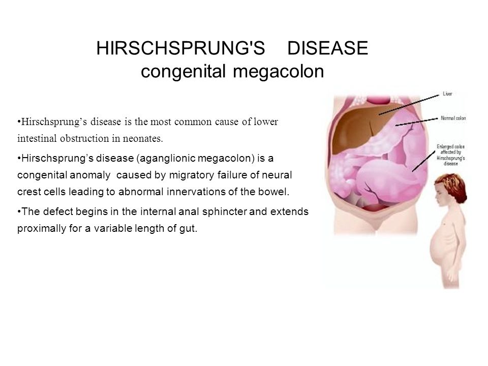
Image taken from: http://slideplayer.com/slide/7541905/24/images/1/HIRSCHSPRUNG+S+DISEASE+congenital+megacolon.jpg


According to the Mayo Clinic (http://www.mayoclinic.org/diseases-conditions/hirschsprung's-disease/home/ovc-20214664), the symptoms of Hirschsprung's Disease are included but not limited to,


"Hirschsprung's (HIRSH-sproongz) disease is a condition that affects the large intestine (colon) and causes problems with passing stool. The condition is present at birth (congenital) as a result of missing nerve cells in the muscles of the baby's colon.

A newborn who has Hirschsprung's disease usually can't have a bowel movement in the days after birth. In mild cases, the condition might not be detected until later in childhood. Uncommonly, Hirschsprung's disease is first diagnosed in adults.

Surgery to bypass or remove the diseased part of the colon is the treatment."


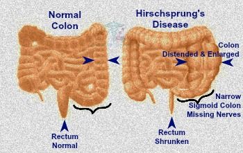
Image taken from: http://www.birth-defect.org/wp-content/uploads/7f5592e639cf5afb772e168b4a153572.jpg


However, even though it is rare or uncommon, adults can very much get it as well.


The symptoms, according to Mayo (http://www.mayoclinic.org/diseases-conditions/hirschsprung's-disease/symptoms-causes/dxc-20214666), are,

"Signs and symptoms of Hirschsprung's disease vary with the severity of the condition. Usually signs and symptoms appear shortly after birth, but sometimes they're not apparent until later in life.

Typically, the most obvious sign is a newborn's failure to have a bowel movement within 48 hours after birth.


Other signs and symptoms in newborns may include:

Swollen belly

Vomiting, including vomiting a green or brown substance

Constipation or gas, which might make a newborn fussy

Diarrhea


In older children/adults, signs and symptoms can include:

Swollen belly

Chronic constipation

Gas

Failure to thrive

Fatigue


It's not clear what causes Hirschsprung's disease. It sometimes occurs in families and might, in some cases, be associated with a genetic mutation.

Hirschsprung's disease occurs when nerve cells in the colon don't form completely. Nerves in the colon control the muscle contractions that move food through the bowels. Without the contractions, stool stays in the large intestine.


Factors that may increase the risk of Hirschsprung's disease include:

Having a sibling who has Hirschsprung's disease. Hirschsprung's disease can be inherited. If you have one child who has the condition, future biological children could be at risk.

Being male. Hirschsprung's disease is more common in males.

Having other inherited conditions. Hirschsprung's disease is associated with certain inherited conditions, such as Down syndrome and other abnormalities present at birth, such as congenital heart disease."


Image taken from: http://www.medindia.net/images/common/patientinfo/950_400/abdominal-x-ray-can-helps-to-diagnose-the-hirschsprung-disease.jpg


According to MedicineNet (http://www.medicinenet.com/hirschsprung_disease/article.htm,

"The cause of Hirschsprung disease is due to nerve cells that are supposed to grow along the intestine and reach the anus, but do not because they stop growing too soon. Hirschsprung disease (HSCR) is a congenital (present at birth) disease of the large intestine or colon. It is one type of birth defect. People with the disease do not have the nerve cells in the intestine required to expel stools from the body normally.

Some people inherit the disease, and others have mutations in several genes. In about 50% of people with Hirschsprung disease, researchers and doctors do not know what genes cause it.

Your primary care doctor will refer you to a specialist in digestive disorders called a gastroenterologist, to diagnose the condition. Diagnosis for Hirschsprung’s is based on a physical exam, medical and family history, symptoms, and tests, for example, a digital exam.
Surgery is the treatment for this life-threatening disease (procedures include pull-through for infants and ostomy (An ostomy refers to the surgically created opening in the body for the discharge of body wastes) for toddles and older children).

After healing from surgery, your child's bowel movements may become normal. But surgery doesn't cure Hirschsprung disease. Some children/adults will have bowel problems – like constipation or fecal incontinence (“accidents”) – off and on throughout their lives.


Symptoms of Hirschsprung disease in toddlers and older children may include:

Not being able to pass stools without enemas or suppositories. An enema involves flushing liquid into the child’s anus using a special wash bottle. A suppository is a pill Placed into the child’s rectum.

Swelling of the abdomen.

Diarrhea, often with blood.

Slow growth.

Intellectual disability"


Image taken from: http://image.slidesharecdn.com/hirschsprungsdisease-140510235224-phpapp01/95/hirschsprungs-disease-3-638.jpg?cb=1399767420


According to Science Direct (http://www.sciencedirect.com/science/article/pii/S2237936315000519,

"Some patients reach adulthood without a diagnosis for this disease. Typically, patients go to the doctor with a long-standing history of constipation requiring frequent laxative use.11 The current frequency of the disease in adults is unknown, especially since HD is an overlooked and misdiagnosed disease in this age group. ...the diagnosis of HD is supported by barium enema studies, anorectal manometry and rectal biopsy.

Imaging studies such as computed tomography (CT) and barium enemas are usually accepted for evaluation of chronic constipation, which is a common disorder in adults. Our patient underwent CT due to the unavailability of a barium enema study, but CT is a more expensive method.

The anorectal manometry, even though not contributing in the present case, is an ancillary test of the utmost importance, since the presence of the rectum-anal reflex in this exam usually rule out the diagnosis of HD.

Several procedures are used to manage this disease after childhood; currently the option of choice is the surgical procedure of Duhamel. Late diagnosis contributes to the need for surgery in more than one surgical time, with ileostomy or colostomy, since the healthy colon is more distended in adolescents and adults compared to neonates and children. Nevertheless, the literature considers as the procedure of choice the Duhamel technique in only one surgical time, which reduces the hospitalization time.

This surgery is considered curative. However, post-operative bowel functioning is not always satisfactory. Enterocolitis, constipation and fecal incontinence represent the main postoperative complications in children. To date, the progression for adolescent or adult patients is not fully clarified yet, due to the small number of reported cases."


Image taken from: https://i.pinimg.com/736x/80/76/5d/80765d68dfe49fd160965c6aa5410100--menu-google-search.jpg


I really hope this will help educate you about Hirschsprung disease. I am still learning about it myself. The research I did says it is more likely to happen with children, but adults I know have it and were just officially diagnosed. After you have a rectal biopsy (because doctors do a biopsy to verify you do have HD), then they might do an anorectal manometry (which is where they stick a balloon up your rectum, kind of like the pelvic floor test). Once all of that has been confirmed, then they might start talking reconstructive pelvic surgery.


I would still advise you to be cautious, do your own research, talk to people who have had it done, and make an informed decision so you will know if this is right for you. Thank you very much to my friend who brought this illness to my attention, because I had no idea this was going on. My heart goes out to all of you who are fighting this on a daily basis.

Tuesday, June 6, 2017

Nissen Fundoplication

A Nissen fundoplication, or laparoscopic Nissen fundoplication when performed via laparoscopic surgery, is a surgical procedure to treat gastroesophageal reflux disease (GERD) and hiatal hernia. In a fundoplication, the gastric fundus (upper part) of the stomach is wrapped, or plicated, around the lower end of the esophagus and stitched in place, reinforcing the closing function of the lower esophageal sphincter. The esophageal hiatus is also narrowed down by sutures to prevent or treat concurrent hiatal hernia, in which the fundus slides up through the enlarged esophageal hiatus of the diaphragm.

In a Nissen fundoplication, also called a complete fundoplication, the fundus is wrapped the entire 360 degrees around the esophagus. In contrast, surgery for achalasia is generally accompanied by either a Dor or Toupet partial fundoplication, which is less likely than a Nissen wrap to aggravate the dysphagia that characterizes achalasia. In a Dor (anterior) fundoplication, the fundus is laid over the top of the esophagus; while in a Toupet (posterior) fundoplication, the fundus is wrapped around the back of the esophagus.

The procedure is now routinely performed laparoscopically. When used to alleviate gastroesophageal reflux symptoms in patients with delayed gastric emptying (gastroparesis), it is frequently combined with modification of the pylorus via pyloromyotomy or pyloroplasty. (You can read that here in another blog article of mine: http://www.emilysstomach.com/2017/06/pyloric-stent-pyloroplasty.html) There is also a new Facebook support group for those who have had it done, and also combat Gastroparesis: GASTROPARESIS AFTER BARIATRIC SURGERY.


This is a completed Nissen Fundoplication in Watercolors:
Source: By Dana Hamers - Own work, CC BY-SA 3.0, https://commons.wikimedia.org/w/index.php?curid=20453728
Source: http://en.wikipedia.org/wiki/Nissen_fundoplication


This is the Nissen Fundoplication Procedure:
Source: Original Artwork by James P. Gray, M.D. 2007 (author)


The doctor that I saw that the Mayo Clinic wanted me to have this surgery done so that it would stop my vomiting. However, he concluded that since I vomit so violently, I would undo the surgery. A mom who reads my blog and has a son with gastroparesis, was nice enough to share his story with me about the Nissen he had done. When my doctor said that it would stop vomiting, he was wrong. His story is below,


"Hi, my 17 year old son had the Nissen procedure last year. He too violently throws up. The surgery went well but he said out of his 33 surgeries it was the most painful after. He went through the recovery process not realizing until about a week after that he could not even swallow water without it coming right back up.

He lost weight quickly and was very dehydrated so at his check up appointment the hospitalized him. The Nissen was too tight and nothing was going to his stomach. The doctor did a scope and stretched the area. That helped for a bit but they had to go back and stretch it a second time.

That was the last stretch they had to do. They said the Nissen would make it almost impossible for him to throw up. Boy were they wrong. He still throws up most every time he eats. By the way he has GP (gastroparesis) and has an electric stimulator in his stomach. Amanda S."


I also know a few people with gastroparesis who have had this surgery done and they are miserable now. They are unable to vomit at all. That worries me, personally, because what if you get food poisoning and need to vomit to get it out? What do you do then?


"Gastroparesis Following Nissen Fundoplication And Hiatal Hernia Repair

Nissen fundoplication with hiatal hernia repair is the most reliable and most effective treatment of GERD or acid reflux disease. The procedure is also very safe with less than 1% complication rate. Gastroparesis or delayed gastric emptying is a poorly understood medical disorder. Gastroparesis results from abnormal gastro-duodenal motility resulting in nausea, vomiting, bloating, epigastric pain and early satiety. Gastroparesis can also contribute to acid reflux disease. GERD is a multi-factorial problem and is closely related to gastric motility. Indeed, gastric fundus compliance, relaxation, food accommodation and luminal pressure affects transient lower esophageal sphincter relaxation, TLESR. TLESR is believed to be the main cause of acid reflux. It is not surprising for gastroparesis patients to suffer from heartburn and other GERD related symptoms. In fact, both GERD and gastroparesis may represent different aspects of the same problem related to esophago-gastro-intestinal dysmotility.

Many GERD patients undergoing Nissen fundoplication and hiatal hernia repair surgery may also have undiagnosed gastroparesis. Around 40% of GERD patients suffer from delayed gastric emptying. Nissen fundoplication increases gastric emptying and is sometimes associated with dumping especially in children. Wrapping the fundus around the esophagus decreases gastric compliance possibly leading to increased gastric emptying. The same mechanism of action may also be at play in the case of sleeve gastrectomy. By resecting the gastric fundus, gastric compliance decreases and emptying increases. Consequently, Nissen surgery improves gastric emptying and it contributes to gastroparesis symptom resolution.

In a minority of patients, Nissen surgery is associated with post-operative gastroparesis symptom development. These patients develop nausea, bloating, and pain in the first few days after Nissen surgery. It is unclear whether the surgery itself causes de novo gastroparesis or if it exacerbates an already existing problem with gastric emptying. It has always been assumed that vagal nerve injury results in gastric stasis and failure of the pylorus to relax. Pyloromyotomy has also been advocated in vagotomy cases. Swanstrom et al published a study in 2009 in Archives of Surgery titled “Outcomes of Nissen Fundoplication in Patients with Gastroesophageal Reflux Disease and Delayed Gastric Emptying”. He recommends the addition of pyloroplasty to Nissen fundoplication in cases of delayed gastric emptying. In my experience, pyloromyotomy, like other drainage procedures, has minimal effect on gastric emptying. Gastric emptying is a highly coordinated myo-electrical process. Many feedback signals, in addition to the vagus nerve, modulate this activity. It is unlikely that post-operative gastroparesis is the sole result of vagal nerve injury.

In 2004, a study published by Masclee et al, in the Annals of Surgery showed that laparoscopic fundoplication increases gastric emptying independent of vagal nerve function. The authors nicely showed that 10% of fundoplication patients developed vagal nerve dysfunction post-operatively without affecting gastric emptying or the efficacy of fundoplication in controlling acid reflux. The etiology of gastric stasis is following Nissen surgery remains unclear. There are no established treatment guidelines for gastroparesis. Medications, gastric pacing and drainage procedures are not effective solutions. Subtotal gastrectomy and gastric bypass are associated with poor outcomes. Longitudinal gastrectomy with or without duodeno-jejunostomy seems to be very effective in curing gastroparesis. I have developed this technique several years ago and I have had the chance to apply it on 4 patients so far. Last year, a young man with gastroparesis following fundoplication surgery at an outside institution presented to my office. I performed a longitudinal gastrectomy while preserving the fundoplication and antrum. His symptoms improved immediately. A post-operative UGI study on day one after surgery showed normal gastric emptying. The patient is 8-month post-op now and still doing great.

In summary, gastroparesis and gastric emptying remain poorly understood. However, a tailored longitudinal gastrectomy, even in the presence of a fundoplication, may be an effective and durable solution for gastroparesis. Additional studies are needed to establish this approach as the standard of care treatment for gastroparesis."
Source: http://houstonheartburn.com/gastroparesis-following-nissen-fundoplication-and-hiatal-hernia-repair/


Source: http://www.laparoscopicsurgeonmumbai.com/precautions-nissen-laparoscopic-fundoplication-surgery-gerd/


WebMD says,

"Surgery Overview

During fundoplication surgery, the upper curve of the stomach (the fundus) is wrapped around the esophagus camera and sewn into place so that the lower portion of the esophagus passes through a small tunnel of stomach muscle. This surgery strengthens the valve between the esophagus and stomach (lower esophageal sphincter), which stops acid from backing up into the esophagus as easily. This allows the esophagus to heal.

This procedure can be done through the abdomen or the chest. The chest approach is often used if a person is overweight or has a short esophagus.

This procedure is often done using a laparoscopic surgical technique. Outcomes of the laparoscopic technique are best when the surgery is done by a surgeon with experience using this procedure.

If a person has a hiatal hernia, which can cause gastroesophageal reflux disease (GERD) camera.gif symptoms, it will also be repaired during this surgery.


What To Expect After Surgery

If open surgery (which requires a large incision) is done, you will most likely spend several days in the hospital. A general anesthetic is used, which means you sleep through the operation. After open surgery, you may need 4 to 6 weeks to get back to work or your normal routine.

If the laparoscopic method is used, you will most likely be in the hospital for only 2 to 3 days. A general anesthetic is used. You will have less pain after surgery, because there is no large incision to heal. After laparoscopic surgery, most people can go back to work or their normal routine in about 2 to 3 weeks, depending on their work.

After either surgery, you may need to change the way you eat. You may need to eat only soft foods until the surgery heals. And you should chew food thoroughly and eat more slowly to give the food time to go down the esophagus.


Why It Is Done

Fundoplication surgery is most often used to treat GERD symptoms that are likely to be caused in part by a hiatal hernia and that have not been well controlled by medicines. The surgery may also be used for some people who do not have a hiatal hernia. Surgery also may be an option when:

Treatment with medicines does not completely relieve your symptoms, and the remaining symptoms are proved to be caused by reflux of stomach juices.

You do not want or, because of side effects, you are unable to take medicines over an extended period of time to control your GERD symptoms, and you are willing to accept the risks of surgery.

You have symptoms that do not adequately improve when treated with medicines. Examples of these symptoms are asthma, hoarseness, or cough along with reflux.


How Well It Works

In most people who have laparoscopic surgery for GERD, the surgery improves symptoms and heals the damage done to the esophagus.

Over time, some people have symptoms come back, have esophagitis come back, need to take medicine for symptoms, or need another operation.

Surgery can cause new and troublesome symptoms. Over time, some people have trouble swallowing, have increased flatulence (gas), and/or have trouble belching.


Risks or complications following fundoplication surgery include:

Difficulty swallowing because the stomach is wrapped too high on the esophagus or is wrapped too tightly.

The esophagus sliding out of the wrapped portion of the stomach so that the valve (lower esophageal sphincter) is no longer supported.
Heartburn that comes back.

Bloating and discomfort from gas buildup because the person is not able to burp.

Excess gas.

Risks of anesthesia.

Risks of major surgery (infection or bleeding).

For some people, the side effects of surgery-bloating caused by gas buildup, swallowing problems, pain at the surgical site-are as bothersome as GERD symptoms. The fundoplication procedure cannot be reversed, and in some cases it may not be possible to relieve the symptoms of these complications, even with a second surgery.


What To Think About

GERD can be annoying and even painful. But it is not a dangerous disease. For any GERD treatment to be worth trying, it needs to be very safe. For many people, especially those who have few problems taking medicine, surgery is not a good choice.

But when fundoplication surgery is successful, it may end the need for long-term treatment with medicine. When you are deciding between surgery and treatment with medicine, weigh the cost, risks, and potential complications of the surgery against the cost and inconvenience of taking medicine.


GERD: Which Treatment Should I Use?

Before surgery, additional tests will usually be done to be sure that surgery is likely to help cure GERD symptoms and to diagnose problems that could be made worse by surgery.

Second surgeries are harder to do, are less successful, and are more risky. So it is extremely important that the first procedure be considered carefully and be done by an experienced surgeon who is more likely to be successful the first time.


Surgery to treat GERD is rarely done on people who:

Are older adults, especially if they have other health problems in addition to GERD.

Have weak squeezing motions (peristalsis) in the esophagus. These motions are important to move food down the esophagus to the stomach. Surgery may make this problem worse, causing food to get stuck in the esophagus.

Have unusual symptoms that might be made worse by surgery.

In special cases, other surgeries such as partial fundoplication or gastropexy may be done instead of fundoplication surgery.


Source: http://www.bariatricinnovationsatl.com/acid-reflux-surgery


References and Citations:

Lundell L, et al. (2007). Seven-year follow-up of a randomized clinical trial comparing proton-pump inhibition with surgical therapy for reflux oesophagitis. British Journal of Surgery, 94(2): 198-203.


Other Works Consulted:

Galmiche J-P, et al. (2011). Laparoscopic antireflux surgery vs esomeprazole treatment for chronic GERD. JAMA, 305(19): 1969-1977.


Credits:

By: Healthwise Staff
Primary Medical ReviewerAdam Husney, MD - Family Medicine
Specialist Medical ReviewerPeter J. Kahrilas, MD - Gastroenterology

Current as of November 20, 2015"


Friday, July 15, 2016

Information about The Vagus Nerve

Dedicated to Cheryl, my inspiration for this article.


Image taken from: http://autismcoach.com/blog-info-articles/the-vagus-nerve-core-piece-of-the-autism-puzzle-part-1-what-is-this-and-why-is-it-important

I know I try not to cite too many articles but I wanted to cite this one. It has really great information about the vagus nerve and what it does for your body. I do not know much about the vagus nerve, so I need help with research about it, hence the quoted articles. I'm curious as to if the vagus nerve, if damaged and causes gastroparesis, might cause other issues as well. It's a question I've had on my mind for a long time. I know some of my friends have heart issues and the vagus nerve runs by the heart. I also know that I get what I call, "gp fog" which could be a result of a damaged vagus nerve. So, I wanted to do some research into it and try to answer some of my questions and hopefully, educate and answer some of your questions as well. Maybe the more exposure we bring to complications like this, the doctors might be able to take the information and do something with it - with research or anything that could help us.


"The vagus nerve provides 75% of all parasympathetic outflow. When the brain triggers parasympathetic activation, the vagus nerve carries the messages to the heart (decreasing the heart rate and blood pressure), to the lungs (to constrict the respiratory passageways), to every organ in the digestive system (to increase motility and blood flow to the digestive tract, to promote defecation), to the kidneys and bladder (to promote urination) and to reproductive organs (to aid in sexual arousal).

2. It communicates messages between the gut and the brain. 80% of the vagus nerve’s fibers (4 out of 5 traffic lanes) deliver information from the enteric nervous system (the second brain in the gut) to the brain.

3. It regulates the muscle movement necessary to keep you breathing. Your brain communicates with your diaphragm via the release of the neurotransmitter acetylcholine from the vagus nerve to keep you breathing. If the vagus nerve stops releasing acetylcholine, you will stop breathing.

4. It helps decrease inflammation. About 15 years ago neurosurgeon Kevin Tracey and his colleagues found that a tiny amount of an anti-inflammatory drug in rats’ brains blocked the production of an inflammatory molecule in the liver and spleen. How did the signal get there? The researchers began cutting nerves one at a time to find the ones responsible for transmitting the anti-inflammatory signal from brain to body. “When we cut the vagus nerve, which runs from the brain stem down to the spleen, the effect was gone,” says Tracey, president and CEO of the Feinstein Institute for Medical Research in Manhasset, N.Y. Later they discovered that stimulating undamaged vagus fibers also had anti-inflammatory effects in animals (without the drug), which they attributed to the release of the neurotransmitter acetylcholine by the vagus nerve.


5. It has profound control over heart rate and blood pressure. For example, patients with heart failure, in which the heart fails to pump enough blood through the body, tend to have less active vagus nerves. Currently multiple studies are underway investigating the effects of vagus stimulation on patients with heart failure and atrial fibrillation (where the heart flutters erratically).

6. It helps improve your mood. Research shows that stimulation of the vagus nerve can be an effective treatment for chronic depression that has failed to respond to other treatments. Electrical stimulation of the vagus through a surgically implanted device has already been approved by the U.S. Food and Drug Administration as a therapy for patients who don’t get relief from existing treatments.

7. It is essential in fear management. Remember that “gut instinct” that tells you when something isn’t right? Turns out that the vagus nerve plays a major role in that. The signals from your gut get sent to the brain via the vagus nerve, and the signals from the brain travel back to the gut, forming a feedback loop. What if this loop was interrupted – wondered the researchers in a new Swiss study – would that affect innate anxiety and conditioned fear? Turns out it does. In test animals, the brain was still able to send signals down to the stomach, but the brain couldn’t receive signals coming up from the stomach. The research showed that those rats weren’t that afraid to begin with (lower level of innate fear), but once they became afraid, they had trouble overcoming this fear even when the danger was no longer present (longer retention of learned fear). This shows that healthy functioning of the vagus nerve helps us bounce back from stressful situations and overcome fear conditioning.

8. It plays a role in learning and memory. The same Swiss study (above) found that the rats without gut instincts transmitting to the brain via the vagus nerve required significantly longer to re-associate previously “dangerous” environment with the new, “safe” and neutral situation. This shows that the vagus nerve facilitates learning and re-wiring, so to speak. “These new findings about the vagus nerve offer exciting possibility for the treatment of post-traumatic stress disorder (PTSD). Stimulation of the vagus nerve might be able to speed up the process by which people with PTSD can learn to reassociate a non-threatening stimuli which triggers anxiety with a neutral and non-traumatic experience”(1). It can also help with healing sexual stress and trauma.

9. It can help relieve cluster headaches. The company electroCore, based in Basking Ridge, N.J., manufactures a small, handheld device that can stimulate the vagus when placed on the throat. The company initially tested the devices to reduce asthma symptoms — relying on the nerve’s anti-inflammatory action. But during testing, patients reported that their headaches were disappearing, says J.P. Errico, CEO of electroCore. Now, the company is investigating the use of an electroCore device to treat chronic cluster headaches." This information was given by: https://www.yogauonline.com/yogau-wellness-blog/vital-vagus-what-vagus-nerve-and-what-does-it-do


The gastric branches (rami gastrici) supply the stomach. The right vagus forms the posterior gastric plexus and the left forms the anterior gastric plexus. The branches lie on the posteroinferior and the anterosuperior surfaces, respectively.

The celiac branches (rami celiaci) are derived mainly from the right vagus nerve. They join the celiac plexus and supply the pancreas, spleen, kidneys, adrenals, and intestine.

The hepatic branches originate from the left vagus. They join the hepatic plexus and through it are distributed to the liver.

Images from: http://autismcoach.com/blog-info-articles/the-vagus-nerve-core-piece-of-the-autism-puzzle-part-1-what-is-this-and-why-is-it-important/


"What happens in the vagus nerve, it turns out, doesn’t stay in the vagus nerve. The longest of the cranial nerves, the vagus nerve is so named because it “wanders” like a vagabond, sending out fibers from your brainstem to your visceral organs. The vagus nerve is literally the captain of your inner nerve center—the parasympathetic nervous system, to be specific. And like a good captain, it does a great job of overseeing a vast range of crucial functions, communicating nerve impulses to every organ in your body. New research has revealed that it may also be the missing link to treating chronic inflammation, and the beginning of an exciting new field of treatment that leaves medications behind. Here are nine facts about this powerful nerve bundle.

1. THE VAGUS NERVE PREVENTS INFLAMMATION.
With a vast network of fibers stationed like spies around all your organs, when the vagus nerve gets wind of the hallmarks of inflammation—cytokines or the inflammatory substance tumor necrosis factor (TNF)—it alerts the brain and elicits anti-inflammatory neurotransmitters via the cholinergic anti-inflammatory pathway. A certain amount of inflammation after injury or illness is normal. But an overabundance is linked to many diseases and conditions, from sepsis to the autoimmune condition rheumatoid arthritis.


2. IT HELPS YOU MAKE MEMORIES.
A University of Virginia study showed success in strengthening memory in rats by stimulating the vagus nerve, which releases the neurotransmitter norepinephrine into the amygdala, consolidating memories. Related studies were done on humans, opening promising treatments for conditions like Alzheimer’s disease.


3. IT HELPS YOU BREATHE.
The neurotransmitter acetylcholine, elicited by the vagus nerve, literally gives you the breath of life by telling your lungs to breathe. It’s one of the reasons that botox—often used cosmetically—can be potentially dangerous, because it interrupts your acetylcholine production. You can, however, also manually stimulate your vagus nerve by doing abdominal breathing or holding your breath for four to eight counts.


4. IT’S INTIMATELY INVOLVED WITH YOUR HEART.
The vagus nerve is responsible for controlling the heart rate via electrical impulses to the sinoatrial node of the heart, where acetylcholine release slows the pulse. The way doctors determine the “tone” or “strength” of your vagus nerve (and your cardiac health) is by measuring the time between your individual heart beats, and then plotting this on a chart over time. This is your 'heart rate variability.'


5. IT INITIATES YOUR BODY’S RELAXATION RESPONSE.
When your ever-vigilant sympathetic nervous system revs up the fight or flight responses—pouring the stress hormone cortisol and adrenaline into your body—the vagus nerve tells your body to chill out by releasing acetylcholine. Its tendrils extend to many organs, acting like fiberoptic cables that send instructions to release enzymes and proteins like prolactin, vasopressin, and oxytocin, which calm you down. People with a stronger vagus response may be more likely to recover more quickly after stress, injury, or illness.


6. IT TRANSLATES BETWEEN YOUR GUT AND YOUR BRAIN.
Your gut uses the vagus nerve like a walkie-talkie to tell your brain how you’re feeling via electric impulses called “action potentials". Your gut feelings are very real.


7. OVERSTIMULATION OF THE VAGUS NERVE IS THE MOST COMMON CAUSE OF FAINTING.
If you tremble or get queasy at the sight of blood or while getting a flu shot, you’re not weak; you’re experiencing “vagal syncope.” Your body, responding to stress, overstimulates the vagus nerve, causing your blood pressure and heart rate to drop. During extreme syncope, blood flow is restricted to your brain, and you lose consciousness. But most of the time you just have to sit or lie down for the symptoms to subside.


8. ELECTRIC STIMULATION OF THE VAGUS NERVE REDUCES INFLAMMATION AND MAY INHIBIT IT ALTOGETHER.
Truly breaking new medical ground, neurosurgeon Kevin Tracey was the first to prove that stimulating the vagus nerve can significantly reduce inflammation. Results on rats were so successful, he reproduced the experiment in humans with stunning results. The creation of implants to stimulate the vagus nerve via electronic implants showed a drastic reduction, and even remission, in rheumatoid arthritis—which has no known cure and is often treated with the toxic cancer drug methotraxate—hemorrhagic shock, and other equally serious inflammatory syndromes.


9. VAGUS NERVE STIMULATION HAS CREATED A NEW FIELD OF MEDICINE.
Spurred on by the success of vagal nerve stimulation to treat inflammation and epilepsy, a burgeoning field of medical study, known as “bioelectronics,” may be the future of medicine. Using implants that deliver electric impulses to various body parts, scientists and doctors hope to treat illness with fewer medications and fewer side effects."
Information from: http://mentalfloss.com/article/65710/9-nervy-facts-about-vagus-nerve


I'm sorry for all of the block quotes. I really don't know much about the vagus nerve, so I wanted to read up on it. I feel like it's something I should be aware of since I have Gastroparesis and a damaged vagus nerve and I wanted to spread the knowledge to you. Feel free to do more research on the matter and if I have left out any important information, let me know. I did not know that the vagus nerve can control your "gut feeling." I'm still learning about gastroparesis and what it is doing to my body. I know a lot of my friends have a hard time remembering things, myself included, and a lot of my friends have heart issues. I wonder if it's all related to the damaged vagus nerve by the stomach. I would love a doctor to do research and confirm what a damaged vagus nerve can do to the whole body, and not just the stomach.

The Mayo Clinic offers treatment for a stimulation of the vagus nerve. Their website says,

"Vagus nerve stimulation is a procedure that involves implantation of a device that stimulates the vagus nerve with electrical impulses.

There's one vagus nerve on each side of your body, running from your brainstem through your neck to your chest and abdomen.

Vagus nerve stimulation is most often used to treat epilepsy when other treatments haven't worked. Vagus nerve stimulation is also a treatment for hard-to-treat depression that hasn't responded to typical therapies.

Researchers are currently studying vagus nerve stimulation as a potential treatment for a variety of conditions, including multiple sclerosis, headache, pain and Alzheimer's disease.

In conventional vagus nerve stimulation, a device is surgically implanted under the skin on your chest, and a wire is threaded under your skin connecting the device to the left vagus nerve. The right vagus nerve is not used because it carries fibers that supply nerves to the heart.

When activated, the device sends electrical signals along the vagus nerve to your brainstem, which then sends signals to certain areas in your brain.

New, noninvasive vagus nerve stimulation devices, which do not require surgical implantation, have been approved for use in Europe to treat epilepsy, depression and pain but have not yet been approved for use in the U.S.

An implantable device that stimulates the right vagus nerve is also under study for the treatment of heart failure."
Source: http://www.mayoclinic.org/tests-procedures/vagus-nerve-stimulation/home/ovc-20167755

I hope one day, there will be some kind of treatment to help those with gastroparesis that includes the vagus nerve. I'm optimistic and I'm hoping there will be research into the matter. If we could repair the vagus nerve in some way, it would save so many lives, or even find treatment to get it to work properly. I'm not going to lose hope.


Source: http://thehigherlearning.com/wp-content/uploads/2015/05/vagus-nerve.jpg


There is an article I wrote on the Vagus Nerve Stimulator, which may help your stomach muscles contract and help with gastric emptying. It can be found here: http://www.emilysstomach.com/2017/06/the-vagus-nerve-stimulator.html

Additionally, I also wrote an article entitled, "The Brain in Your Gut" which the deals with the Vagus Nerve and Serotonin being made in your stomach. It can be found here: http://www.emilysstomach.com/2017/09/the-brain-in-your-gut.html


Source: https://feedyeti.com/hashtag.php?q=Gastroparesis


According to Psychology Spot,

"Vagus nerve and anxiety: Everything You Need to Know


The vagus nerve is the tenth of twelve pairs of cranial nerves and is the longest in the body. In fact, the word vagus means 'vagabond' in Latin, and perfectly illustrates the path of this nerve extending through various organs of the body.

The vagus nerve is born in the cranial box, exactly in the spinal cord, and falls into the neck developing on two branches and reaching the abdomen passing through the various organs along the path.

The vagus nerve intervenes in the sensitivity of the respiratory mucous membranes and transmits the rhythm, strength and frequency of breathing. It affects the pharynx, the larynx, the esophagus, the trachea and the bronchi, as well as administering nerve fibers to the heart, stomach, pancreas and liver. But it also carries out the inverse mission; that is, it receives signals from the internal organs and sends them to the brain to be processed.

Although perhaps the most interesting thing is the relationship between the vagus nerve and anxiety as it also transmits signals of nervousness or calm, anger or relaxation.


To understand the link between the vagus nerve and anxiety we need to understand that the nervous system is made up of two 'opposite' systems that constantly send information to the brain.

The sympathetic nervous system prepares us for action, so it mainly feeds hormones like adrenaline and cortisol. The parasympathetic nervous system intervenes in rest and relaxation.

In practice, both systems work as accelerator and decelerator. The sympathetic nervous system accelerates and activates us as the parasympathetic nervous system helps us relax and reduce the speed, so it uses neurotransmitters such as acetylcholine, which decreases the heart rate and blood pressure so that the organs work slower.


The functions of the vagus nerve

The vagus nerve controls the parasympathetic system. It intervenes in many functions, from mouth movements to heartbeat, and likewise, when affected ​​it can cause various symptoms. Some of the vagus nerve functions in our body are:

– It helps regulate heartbeat, controls muscle movements and maintains the pace of breathing.

– It maintains the functioning of the digestive tract, allowing the contraction of the stomach and intestine muscles to digest food.

– Facilitates relaxation after a stressful situation or indicates that we are in danger and we do not have to lower the guard.

– Send sensory information to the brain about organ status.


Vagus Nerve and Anxiety

When we are subjected to stressful situations, the sympathetic nervous system is activated. If the tension persists and we cannot turn off the physiological response that triggers it, it won’t pass much time before problems appear. At brain level, this involves the activation of two pathways: the hypothalamus-pituitary-adrenal axis and the brain-intestine axis.

The brain responds to stress and anxiety by increasing the production of hormones (CRFs) that travel from the hypothalamus to the pituitary gland where they induce the release of another hormone (ACTH), which in turn travels through the bloodstream to the adrenal glands to stimulate cortisol and adrenaline induction, which act as immune system suppressors and inflammatory precursors, which is why when we feel stressed and anxious we get ill easily and, ultimately, we can end up suffering from depression, a disorder which has been linked to an inflammatory brain response.

And as if that were not enough, chronic stress and anxiety cause an increase in glutamate in the brain, a neurotransmitter that, when produced in excess, causes migraine, depression and anxiety. In addition, a high level of cortisol reduces the volume of the hippocampus, the part of the brain responsible for the formation of new memories.

The involvement of the vagus nerve will lead to symptoms such as dizziness, gastrointestinal problems, arrhythmias, difficulty in breathing, and disproportionate emotional responses. In fact, as the vagus nerve is unable to activate the relaxation signal, the sympathetic nervous system keeps active, this will cause the person to respond impulsively and suffer from anxiety.

It is also curious that a study developed at the University of Miami found that the vagal tone is transmitted from mother to child. Women suffering from anxiety, depression or experiencing much anger during pregnancy had a lower vagal activity and their children also exhibited low vagal activity and lower levels of dopamine and serotonin.


Three Vagal Stimulation Techniques: How to Take Care of the Vagus Nerve?

The vagal tone is an internal biological process that represents the activity of the vagus nerve. The increase in vagal tone activates the parasympathetic nervous system, which means that we can relax more quickly after a stressful situation and this will have a positive impact on our emotional balance and on health in general.

Exist various vagus nerve stimulation techniques:

1. Exposure to cold

It has been seen that exposure to cold activates the vagus nerve because it stimulates the cholinergic neurons crossing these innervations. In fact, an investigation conducted at the University of Oulu has revealed that regular exposure to cold helps to reduce the fight-flight response that launches the sympathetic nervous system.

It can be enough a cold shower of 30 seconds a day or a cold towel on the face. There are also those who lie down on the belly putting a cube of ice on the nape. Others prefer to drink quickly a glass of cold water.

2. Diaphragmatic breathing

Most people inhale air between 10 and 14 times per minute, which means they have a superficial breathing. The ideal would be to inhale air 6 times per minute. Therefore, another very effective vagal stimulation technique consists in breathing deeply.

The diaphragmatic breathing in particular activates the vagus nerve and the brain interprets it as it is necessary to calm down, even if the nerve has not given that order specifically. The mechanism is the same for which, if you close your eyes and make taps with your fingers on your eyelids, you will perceive short flashes of light because the brain interprets them so.

With diaphragmatic breathing, we make a deeper breathing that brings air into the lower part of the chest, using the diaphragm correctly and promoting relaxation.

3. Meditation, yoga and tai-chi

Meditation can increase the vagal tone. This has been demonstrated by researchers of the Oregon University who have seen that only five days of mindfulness meditation promote positive feelings towards oneself that cause the vagus nerve activation, while modulating the activity of the parasympathetic nervous system, a much better result than conventional relaxation techniques.

Even practices such as yoga and tai-chi are ideal for stimulating the vagus nerve. A study at Boston University has revealed that yoga increases GABA neurotransmitters, which promote the feeling of calm and serenity by helping to combat anxiety and stress. The tai-chi, on the other hand, is able to balance heart rate, which means it stimulates vagal modulation, according to researchers at the National Yang-Ming University School of Medicine.


Sources:
Streeter, C. C. et. Al. (2012) Effects of yoga on the autonomic nervous system, gamma-aminobutyric-acid, and allostasis in epilepsy, depression, and post-traumatic stress disorder. Medical Hypotheses; 78(5): 571-579.
Tang, Y. et. Al. (2009) Central and autonomic nervous system interaction is altered by short-term meditation. PNAS; 106(22): 8865–8870.
Mäkinen, T.M. et. Al. (2008) Autonomic nervous function during whole-body cold exposure before and after cold acclimation. Aviat Space Environ Med; 79(9): 875-882.
Fiel, T. et. Al. (2003) Pregnancy anxiety and comorbid depression and anger: Effects on the fetus and neonate. Depression and Anxiety; 17(3): 140–151.
Wan-an, L. & Cheng-Deng, K (2003) The Effect of Tai Chi Chuan on the Autonomic Nervous Modulation in Older Persons. Med Sci Sports Exerc; 35(12): 1972-1976."


Source: https://www.theodysseyonline.com/vagus-nerve-dysfunction-symptoms

Thursday, May 9, 2013

pH Probe Test - Mayo Visit Day 1

Well, I went to Mayo this morning and had the pH probe test done. I sat down and the nurse explained the test to me. The probe will test my vomit for bile, stomach acid, regurgitation, etc. This test will help the doctor determine how best to treat me. She had me sit facing her while she inserted the small catheter into my nose, down my throat, and into my stomach. She didn't use any numbing agents or anything before she put the catheter in. My eyes watered like crazy and she gave me water to sip on while she was inserting the catheter, to make it easier for me not to gag.

Once the catheter was in, the nurse secured it with tape. However, I do not believe it's secured that well because the tube tends to move in and out of my nose with every bump in the car, swallow, or movement. It's going to take some getting used to.


Illustration of where the pH Probe goes.


The nurse told me that I needed to take in three meals to ensure an accurate test. I'm not able to eat that much on the best of days, so this is going to be a real challenge for me.

She gave me a receiver that I have to wear, much like the Bravo Test that I had before. The receiver has three buttons that I need to press when I take my Aciphex, if I have heartburn, and if I experience regurgitation. I have to press another button when I eat and when I finish eating as well as when I lay down or up and about. The catheter is attached to this receiver, so I can't leave it on the bed or anything. I MUST carry it with me.


The receiver I have to carry with me.


So, I will wear the pH probe until tomorrow morning. I've got to eat in the morning, which will be difficult because that's when I'm the most sick. The nurse said she'd take it out before I see the doctor in the morning for the results.

My nose is really irritated and I keep sneezing. The tube keeps gagging me but other than that, it's not to bad. I prefer the Bravo Test though.


My pH probe!